


 Foundational characteristics of cancer include proliferation, angiogenesis, migration, evasion of apoptosis, and cellular immortality. Find key markers for these cellular processes and antibodies to detect them.
 The SUMOplot™ Analysis Program predicts and scores sumoylation sites in your protein. SUMOylation is a post-translational modification involved in various cellular processes, such as nuclear-cytosolic transport, transcriptional regulation, apoptosis, protein stability, response to stress, and progression through the cell cycle.
 The Autophagy Receptor Motif Plotter predicts and scores autophagy receptor binding sites in your protein. Identifying proteins connected to this pathway is critical to understanding the role of autophagy in physiological as well as pathological processes such as development, differentiation, neurodegenerative diseases, stress, infection, and cancer.
Anti-NHE9 Antibody
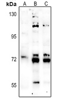
Rabbit polyclonal antibody to NHE9
- SPECIFICATION
- CITATIONS
- PROTOCOLS
- BACKGROUND

Application
| WB, IP |
|---|---|
| Primary Accession | Q8IVB4 |
| Other Accession | Q8BZ00 |
| Reactivity | Human, Mouse, Rat, Monkey, Dog |
| Host | Rabbit |
| Clonality | Polyclonal |
| Calculated MW | 72565 Da |
| Gene ID | 285195 |
|---|---|
| Other Names | NHE9; Sodium/hydrogen exchanger 9; Na(+)/H(+) exchanger 9; NHE-9; Solute carrier family 9 member 9 |
| Target/Specificity | KLH-conjugated synthetic peptide encompassing a sequence within the center region of human NHE9. The exact sequence is proprietary. |
| Dilution | WB~~WB (1/500 - 1/1000), IP (1/10 - 1/100) IP~~N/A |
| Format | Liquid in 0.42% Potassium phosphate, 0.87% Sodium chloride, pH 7.3, 30% glycerol, and 0.09% (W/V) sodium azide. |
| Storage | Store at -20 °C.Stable for 12 months from date of receipt |
| Name | SLC9A9 (HGNC:20653) |
|---|---|
| Synonyms | NHE9 |
| Function | Endosomal Na(+), K(+)/H(+) antiporter. Mediates the electroneutral exchange of endosomal luminal H(+) for a cytosolic Na(+) or K(+) (Probable). By facilitating proton efflux, SLC9A9 counteracts the acidity generated by vacuolar (V)-ATPase, thereby limiting luminal acidification. Regulates organellar pH and consequently, e.g., endosome maturation and endocytic trafficking of plasma membrane receptors and neurotransporters (PubMed:15522866, PubMed:24065030, PubMed:28130443). Promotes the recycling of transferrin receptors back to the cell surface to facilitate additional iron uptake in the brain (PubMed:28130443). Regulates synaptic transmission by regulating the luminal pH of axonal endosomes (By similarity). Regulates phagosome lumenal pH, thus affecting phagosome maturation, and consequently, microbicidal activity in macrophages (By similarity). Can also be active at the cell surface of specialized cells, e.g., in the inner ear hair bundles uses the high K(+) of the endolymph to regulate intracelular pH (By similarity). |
| Cellular Location | Late endosome membrane; Multi-pass membrane protein {ECO:0000250|UniProtKB:F7B113}. Early endosome membrane; Multi-pass membrane protein {ECO:0000250|UniProtKB:F7B113}. Recycling endosome membrane; Multi-pass membrane protein {ECO:0000250|UniProtKB:F7B113}. Cell membrane {ECO:0000250|UniProtKB:Q8BZ00}; Multi-pass membrane protein {ECO:0000250|UniProtKB:F7B113}. Cytoplasmic vesicle, phagosome membrane {ECO:0000250|UniProtKB:Q8BZ00}; Multi-pass membrane protein {ECO:0000250|UniProtKB:F7B113}. Note=Localized to the plasma membrane in inner ear hair cell bundle. {ECO:0000250|UniProtKB:Q8BZ00} |
| Tissue Location | Ubiquitously expressed in all tissues tested. Expressed at highest levels in heart and skeletal muscle, followed by placenta, kidney, and liver. Expressed in the brain, in the medulla and spinal cord. |

Thousands of laboratories across the world have published research that depended on the performance of antibodies from Abcepta to advance their research. Check out links to articles that cite our products in major peer-reviewed journals, organized by research category.
info@abcepta.com, and receive a free "I Love Antibodies" mug.

Provided below are standard protocols that you may find useful for product applications.
Background
KLH-conjugated synthetic peptide encompassing a sequence within the center region of human NHE9. The exact sequence is proprietary.
If you have used an Abcepta product and would like to share how it has performed, please click on the "Submit Review" button and provide the requested information. Our staff will examine and post your review and contact you if needed.
If you have any additional inquiries please email technical services at tech@abcepta.com.
Ordering Information
Other Products
Shipping Information
