


 Foundational characteristics of cancer include proliferation, angiogenesis, migration, evasion of apoptosis, and cellular immortality. Find key markers for these cellular processes and antibodies to detect them.
 The SUMOplot™ Analysis Program predicts and scores sumoylation sites in your protein. SUMOylation is a post-translational modification involved in various cellular processes, such as nuclear-cytosolic transport, transcriptional regulation, apoptosis, protein stability, response to stress, and progression through the cell cycle.
 The Autophagy Receptor Motif Plotter predicts and scores autophagy receptor binding sites in your protein. Identifying proteins connected to this pathway is critical to understanding the role of autophagy in physiological as well as pathological processes such as development, differentiation, neurodegenerative diseases, stress, infection, and cancer.
Anti-Recoverin Antibody
Rabbit polyclonal antibody to Recoverin
- SPECIFICATION
- CITATIONS
- PROTOCOLS
- BACKGROUND

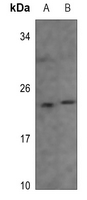
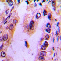
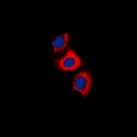
Application
| WB, IF/IC, IHC |
|---|---|
| Primary Accession | P35243 |
| Other Accession | P34057 |
| Reactivity | Human, Mouse, Rat, Pig |
| Host | Rabbit |
| Clonality | Polyclonal |
| Calculated MW | 23130 Da |
| Gene ID | 5957 |
|---|---|
| Other Names | RCV1; Recoverin; Cancer-associated retinopathy protein; Protein CAR |
| Target/Specificity | KLH-conjugated synthetic peptide encompassing a sequence within the center region of human Recoverin. The exact sequence is proprietary. |
| Dilution | WB~~WB (1/500 - 1/1000), IH (1/100 - 1/200), IF/IC (1/100 - 1/500) IF/IC~~N/A IHC~~1:100~500 |
| Format | Liquid in 0.42% Potassium phosphate, 0.87% Sodium chloride, pH 7.3, 30% glycerol, and 0.09% (W/V) sodium azide. |
| Storage | Store at -20 °C.Stable for 12 months from date of receipt |
| Name | RCVRN |
|---|---|
| Synonyms | RCV1 |
| Function | Acts as a calcium sensor and regulates phototransduction of cone and rod photoreceptor cells (By similarity). Modulates light sensitivity of cone photoreceptor in dark and dim conditions (By similarity). In response to high Ca(2+) levels induced by low light levels, prolongs RHO/rhodopsin activation in rod photoreceptor cells by binding to and inhibiting GRK1-mediated phosphorylation of RHO/rhodopsin (By similarity). Plays a role in scotopic vision/enhances vision in dim light by enhancing signal transfer between rod photoreceptors and rod bipolar cells (By similarity). Improves rod photoreceptor sensitivity in dim light and mediates response of rod photoreceptors to facilitate detection of change and motion in bright light (By similarity). |
| Cellular Location | Photoreceptor inner segment {ECO:0000250|UniProtKB:P34057}. Cell projection, cilium, photoreceptor outer segment {ECO:0000250|UniProtKB:P34057}. Photoreceptor outer segment membrane {ECO:0000250|UniProtKB:P21457}; Lipid-anchor {ECO:0000250|UniProtKB:P21457}; Cytoplasmic side {ECO:0000250|UniProtKB:P21457}. Perikaryon {ECO:0000250|UniProtKB:P34057}. Note=Primarily expressed in the inner segments of light-adapted rod photoreceptors, approximately 10% of which translocates from photoreceptor outer segments upon light stimulation (By similarity). Targeting of myristoylated protein to rod photoreceptor outer segments is calcium dependent (By similarity) {ECO:0000250|UniProtKB:P21457, ECO:0000250|UniProtKB:P34057} |
| Tissue Location | Retina and pineal gland. |

Thousands of laboratories across the world have published research that depended on the performance of antibodies from Abcepta to advance their research. Check out links to articles that cite our products in major peer-reviewed journals, organized by research category.
info@abcepta.com, and receive a free "I Love Antibodies" mug.

Provided below are standard protocols that you may find useful for product applications.
Background
KLH-conjugated synthetic peptide encompassing a sequence within the center region of human Recoverin. The exact sequence is proprietary.
If you have used an Abcepta product and would like to share how it has performed, please click on the "Submit Review" button and provide the requested information. Our staff will examine and post your review and contact you if needed.
If you have any additional inquiries please email technical services at tech@abcepta.com.
Ordering Information
Other Products
Shipping Information
