


 Foundational characteristics of cancer include proliferation, angiogenesis, migration, evasion of apoptosis, and cellular immortality. Find key markers for these cellular processes and antibodies to detect them.
 The SUMOplot™ Analysis Program predicts and scores sumoylation sites in your protein. SUMOylation is a post-translational modification involved in various cellular processes, such as nuclear-cytosolic transport, transcriptional regulation, apoptosis, protein stability, response to stress, and progression through the cell cycle.
 The Autophagy Receptor Motif Plotter predicts and scores autophagy receptor binding sites in your protein. Identifying proteins connected to this pathway is critical to understanding the role of autophagy in physiological as well as pathological processes such as development, differentiation, neurodegenerative diseases, stress, infection, and cancer.
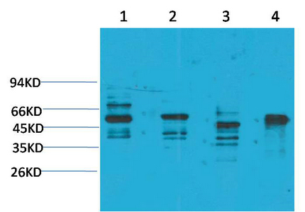
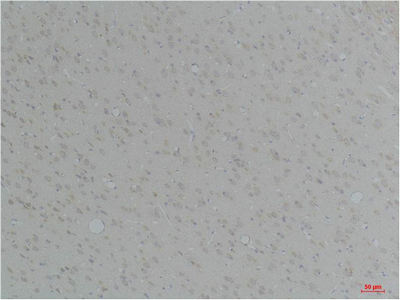
KCNN4 (SK4) Polyclonal Antibody
- SPECIFICATION
- CITATIONS
- PROTOCOLS
- BACKGROUND

Application
| WB, IHC-P |
|---|---|
| Primary Accession | O15554 |
| Reactivity | Human, Rat, Mouse |
| Host | Rabbit |
| Clonality | Polyclonal |
| Calculated MW | 47696 Da |
| Gene ID | 3783 |
|---|---|
| Other Names | KCNN4; IK1; IKCA1; KCA4; SK4; Intermediate conductance calcium-activated potassium channel protein 4; SK4; SKCa 4; SKCa4; IKCa1; IK1; KCa3.1; KCa4; Putative Gardos channel |
| Dilution | WB~~WB 1:1000-2000, IHC 1:50-100 IHC-P~~N/A |
| Format | Liquid in PBS containing 50% glycerol, 0.5% BSA and 0.09% (W/V) sodium azide. |
| Storage Conditions | -20℃ |
| Name | KCNN4 (HGNC:6293) |
|---|---|
| Synonyms | IK1, IKCA1, KCA4, SK4 |
| Function | Intermediate conductance calcium-activated potassium channel that mediates the voltage-independent transmembrane transfer of potassium across the cell membrane through a constitutive interaction with calmodulin which binds the intracellular calcium allowing its opening (PubMed:10026195, PubMed:10961988, PubMed:11425865, PubMed:15831468, PubMed:17157250, PubMed:18796614, PubMed:26148990, PubMed:9326665, PubMed:9380751, PubMed:9407042). The current is characterized by a voltage-independent activation, an intracellular calcium concentration increase-dependent activation and a single- channel conductance of about 25 picosiemens (PubMed:9326665, PubMed:9380751, PubMed:9407042). Also presents an inwardly rectifying current, thus reducing its already small outward conductance of potassium ions, which is particularly the case when the membrane potential displays positive values, above + 20 mV (PubMed:9326665, PubMed:9380751, PubMed:9407042). Controls calcium influx during vascular contractility by being responsible of membrane hyperpolarization induced by vasoactive factors in proliferative vascular smooth muscle cell types (By similarity). Following calcium influx, the consecutive activation of KCNN4 channel leads to a hyperpolarization of the cell membrane potential and hence an increase of the electrical driving force for further calcium influx promoting sustained calcium entry in response to stimulation with chemotactic peptides (PubMed:26418693). Required for maximal calcium influx and proliferation during the reactivation of naive T-cells (PubMed:17157250, PubMed:18796614). Plays a role in the late stages of EGF-induced macropinocytosis through activation by PI(3)P (PubMed:24591580). |
| Cellular Location | Cell membrane; Multi-pass membrane protein. Cell projection, ruffle membrane. Note=Targeted to membrane ruffles after EGF stimulation. |
| Tissue Location | Widely expressed in non-excitable tissues. |

Thousands of laboratories across the world have published research that depended on the performance of antibodies from Abcepta to advance their research. Check out links to articles that cite our products in major peer-reviewed journals, organized by research category.
info@abcepta.com, and receive a free "I Love Antibodies" mug.

Provided below are standard protocols that you may find useful for product applications.
Background
Forms a voltage-independent potassium channel that is activated by intracellular calcium (PubMed:26148990). Activation is followed by membrane hyperpolarization which promotes calcium influx. Required for maximal calcium influx and proliferation during the reactivation of naive T-cells (PubMed:17157250, PubMed:18796614). Plays a role in the late stages of EGF-induced macropinocytosis (PubMed:24591580).
If you have used an Abcepta product and would like to share how it has performed, please click on the "Submit Review" button and provide the requested information. Our staff will examine and post your review and contact you if needed.
If you have any additional inquiries please email technical services at tech@abcepta.com.
Ordering Information
Other Products
Shipping Information
