


 Foundational characteristics of cancer include proliferation, angiogenesis, migration, evasion of apoptosis, and cellular immortality. Find key markers for these cellular processes and antibodies to detect them.
 The SUMOplot™ Analysis Program predicts and scores sumoylation sites in your protein. SUMOylation is a post-translational modification involved in various cellular processes, such as nuclear-cytosolic transport, transcriptional regulation, apoptosis, protein stability, response to stress, and progression through the cell cycle.
 The Autophagy Receptor Motif Plotter predicts and scores autophagy receptor binding sites in your protein. Identifying proteins connected to this pathway is critical to understanding the role of autophagy in physiological as well as pathological processes such as development, differentiation, neurodegenerative diseases, stress, infection, and cancer.
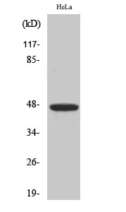
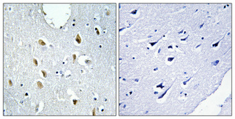
IRF-4 Polyclonal Antibody
- SPECIFICATION
- CITATIONS
- PROTOCOLS
- BACKGROUND

Application
| WB, IHC-P, IF |
|---|---|
| Primary Accession | Q15306 |
| Reactivity | Human, Mouse |
| Host | Rabbit |
| Clonality | Polyclonal |
| Calculated MW | 51772 Da |
| Gene ID | 3662 |
|---|---|
| Other Names | IRF4; MUM1; Interferon regulatory factor 4; IRF-4; Lymphocyte-specific interferon regulatory factor; LSIRF; Multiple myeloma oncogene 1; NF-EM5 |
| Dilution | WB~~Western Blot: 1/500 - 1/2000. Immunohistochemistry: 1/100 - 1/300. Immunofluorescence: 1/200 - 1/1000. ELISA: 1/20000. Not yet tested in other applications. IHC-P~~N/A IF~~1:50~200 |
| Format | Liquid in PBS containing 50% glycerol, 0.5% BSA and 0.09% (W/V) sodium azide. |
| Storage Conditions | -20℃ |
| Name | IRF4 {ECO:0000303|PubMed:15489334, ECO:0000303|PubMed:8921401} |
|---|---|
| Function | Transcriptional activator. Binds to the interferon-stimulated response element (ISRE) of the MHC class I promoter. Binds the immunoglobulin lambda light chain enhancer, together with PU.1. Probably plays a role in ISRE-targeted signal transduction mechanisms specific to lymphoid cells. Involved in CD8(+) dendritic cell differentiation by forming a complex with the BATF-JUNB heterodimer in immune cells, leading to recognition of AICE sequence (5'-TGAnTCA/GAAA- 3'), an immune-specific regulatory element, followed by cooperative binding of BATF and IRF4 and activation of genes. |
| Cellular Location | Nucleus. Cytoplasm |
| Tissue Location | Lymphoid cells. |

Thousands of laboratories across the world have published research that depended on the performance of antibodies from Abcepta to advance their research. Check out links to articles that cite our products in major peer-reviewed journals, organized by research category.
info@abcepta.com, and receive a free "I Love Antibodies" mug.

Provided below are standard protocols that you may find useful for product applications.
Background
Transcriptional activator. Binds to the interferon- stimulated response element (ISRE) of the MHC class I promoter. Binds the immunoglobulin lambda light chain enhancer, together with PU.1. Probably plays a role in ISRE-targeted signal transduction mechanisms specific to lymphoid cells. Involved in CD8(+) dendritic cell differentiation by forming a complex with the BATF-JUNB heterodimer in immune cells, leading to recognition of AICE sequence (5'-TGAnTCA/GAAA-3'), an immune-specific regulatory element, followed by cooperative binding of BATF and IRF4 and activation of genes (By similarity).
If you have used an Abcepta product and would like to share how it has performed, please click on the "Submit Review" button and provide the requested information. Our staff will examine and post your review and contact you if needed.
If you have any additional inquiries please email technical services at tech@abcepta.com.
Ordering Information
Other Products
Shipping Information
