


 Foundational characteristics of cancer include proliferation, angiogenesis, migration, evasion of apoptosis, and cellular immortality. Find key markers for these cellular processes and antibodies to detect them.
 The SUMOplot™ Analysis Program predicts and scores sumoylation sites in your protein. SUMOylation is a post-translational modification involved in various cellular processes, such as nuclear-cytosolic transport, transcriptional regulation, apoptosis, protein stability, response to stress, and progression through the cell cycle.
 The Autophagy Receptor Motif Plotter predicts and scores autophagy receptor binding sites in your protein. Identifying proteins connected to this pathway is critical to understanding the role of autophagy in physiological as well as pathological processes such as development, differentiation, neurodegenerative diseases, stress, infection, and cancer.
MRTF-A Polyclonal Antibody
- SPECIFICATION
- CITATIONS
- PROTOCOLS
- BACKGROUND

Application
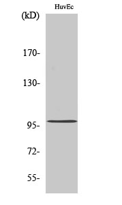
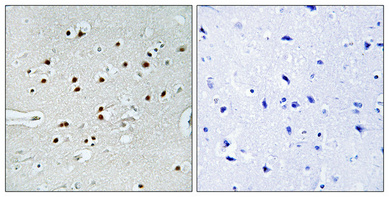
| WB, IHC-P |
|---|---|
| Primary Accession | Q969V6 |
| Reactivity | Human, Mouse |
| Host | Rabbit |
| Clonality | Polyclonal |
| Calculated MW | 98919 Da |
| Gene ID | 57591 |
|---|---|
| Other Names | MKL1; KIAA1438; MAL; MKL/myocardin-like protein 1; Megakaryoblastic leukemia 1 protein; Megakaryocytic acute leukemia protein; Myocardin-related transcription factor A; MRTF-A |
| Dilution | WB~~Western Blot: 1/500 - 1/2000. Immunohistochemistry: 1/100 - 1/300. ELISA: 1/5000. Not yet tested in other applications. IHC-P~~N/A |
| Format | Liquid in PBS containing 50% glycerol, 0.5% BSA and 0.09% (W/V) sodium azide. |
| Storage Conditions | -20℃ |
| Name | MRTFA (HGNC:14334) |
|---|---|
| Function | Transcription coactivator that associates with the serum response factor (SRF) transcription factor to control expression of genes regulating the cytoskeleton during development, morphogenesis and cell migration (PubMed:26224645). The SRF-MRTFA complex activity responds to Rho GTPase-induced changes in cellular globular actin (G- actin) concentration, thereby coupling cytoskeletal gene expression to cytoskeletal dynamics. MRTFA binds G-actin via its RPEL repeats, regulating activity of the MRTFA-SRF complex. Activity is also regulated by filamentous actin (F-actin) in the nucleus. |
| Cellular Location | Cytoplasm. Nucleus Note=Subcellular location is tightly regulated by actin both in cytoplasm and nucleus: high levels of G-actin in the nucleus observed during serum deprivation lead to low levels of nuclear MRTFA, while reduced levels of nuclear G-actin result in accumulation of MRTFA in the nucleus (By similarity). G-actin-binding in the cytoplasm inhibits nuclear import by masking the nuclear localization signal (NLS) (By similarity). In contrast, binding to nuclear globular actin (G-actin) promotes nuclear export to the cytoplasm (By similarity). Nuclear localization is regulated by MICAL2, which mediates depolymerization of nuclear actin, which decreases nuclear G-actin pool, thereby promoting retention of MRTFA in the nucleus and subsequent formation of an active complex with SRF (PubMed:24440334). Nuclear import is mediated by importins KPNA4 and KPNB1 (By similarity) {ECO:0000250|UniProtKB:Q8K4J6, ECO:0000269|PubMed:24440334} |
| Tissue Location | Ubiquitously expressed, has been detected in lung, placenta, small intestine, liver, kidney, spleen, thymus, colon, muscle, heart and brain (PubMed:11344311). Expressed in peripheral blood mononuclear cells (at protein level) (PubMed:26224645) |

Thousands of laboratories across the world have published research that depended on the performance of antibodies from Abcepta to advance their research. Check out links to articles that cite our products in major peer-reviewed journals, organized by research category.
info@abcepta.com, and receive a free "I Love Antibodies" mug.

Provided below are standard protocols that you may find useful for product applications.
Background
Transcription coactivator that associates with the serum response factor (SRF) transcription factor to control expression of genes regulating the cytoskeleton during development, morphogenesis and cell migration. The SRF-MRTFA complex activity responds to Rho GTPase-induced changes in cellular globular actin (G-actin) concentration, thereby coupling cytoskeletal gene expression to cytoskeletal dynamics. MRTFA binds G-actin via its RPEL repeats, regulating activity of the MRTFA-SRF complex. Activity is also regulated by filamentous actin (F-actin) in the nucleus.
If you have used an Abcepta product and would like to share how it has performed, please click on the "Submit Review" button and provide the requested information. Our staff will examine and post your review and contact you if needed.
If you have any additional inquiries please email technical services at tech@abcepta.com.
Ordering Information
Shipping Information
