


 Foundational characteristics of cancer include proliferation, angiogenesis, migration, evasion of apoptosis, and cellular immortality. Find key markers for these cellular processes and antibodies to detect them.
 The SUMOplot™ Analysis Program predicts and scores sumoylation sites in your protein. SUMOylation is a post-translational modification involved in various cellular processes, such as nuclear-cytosolic transport, transcriptional regulation, apoptosis, protein stability, response to stress, and progression through the cell cycle.
 The Autophagy Receptor Motif Plotter predicts and scores autophagy receptor binding sites in your protein. Identifying proteins connected to this pathway is critical to understanding the role of autophagy in physiological as well as pathological processes such as development, differentiation, neurodegenerative diseases, stress, infection, and cancer.
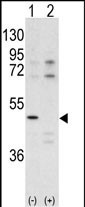
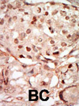
CDK10 Antibody (N-term)
Purified Rabbit Polyclonal Antibody (Pab)
- SPECIFICATION
- CITATIONS: 4
- PROTOCOLS
- BACKGROUND

Application
| IHC-P, WB, E |
|---|---|
| Primary Accession | Q15131 |
| Other Accession | Q3UMM4 |
| Reactivity | Human |
| Predicted | Mouse |
| Host | Rabbit |
| Clonality | Polyclonal |
| Isotype | Rabbit IgG |
| Calculated MW | 41038 Da |
| Antigen Region | 1-32 aa |
| Gene ID | 8558 |
|---|---|
| Other Names | Cyclin-dependent kinase 10, Cell division protein kinase 10, Serine/threonine-protein kinase PISSLRE, CDK10 |
| Target/Specificity | This CDK10 antibody is generated from rabbits immunized with a KLH conjugated synthetic peptide between 1-32 amino acids from the N-terminal region of human CDK10. |
| Dilution | IHC-P~~1:50~100 WB~~1:1000 E~~Use at an assay dependent concentration. |
| Format | Purified polyclonal antibody supplied in PBS with 0.09% (W/V) sodium azide. This antibody is prepared by Saturated Ammonium Sulfate (SAS) precipitation followed by dialysis against PBS. |
| Storage | Maintain refrigerated at 2-8°C for up to 2 weeks. For long term storage store at -20°C in small aliquots to prevent freeze-thaw cycles. |
| Precautions | CDK10 Antibody (N-term) is for research use only and not for use in diagnostic or therapeutic procedures. |
| Name | CDK10 |
|---|---|
| Function | Cyclin-dependent kinase that phosphorylates the transcription factor ETS2 (in vitro) and positively controls its proteasomal degradation (in cells) (PubMed:24218572). Involved in the regulation of actin cytoskeleton organization through the phosphorylation of actin dynamics regulators such as PKN2. Is a negative regulator of ciliogenesis through phosphorylation of PKN2 and promotion of RhoA signaling (PubMed:27104747). |
| Cellular Location | Cytoplasm, cytoskeleton, cilium basal body |

Provided below are standard protocols that you may find useful for product applications.
Background
CDK10 belongs to the CDK subfamily of the Ser/Thr protein kinase family. The CDK subfamily members are highly similar to the gene products of S. cerevisiae cdc28, and S. pombe cdc2, and are known to be essential for cell cycle progression. This kinase has been shown to play a role in cellular proliferation. Its function is limited to cell cycle G2-M phase.
References
Crawford, J., et al., Genomics 56(1):90-97 (1999).
Brambilla, R., et al., Oncogene 9(10):3037-3041 (1994).
Grana, X., et al., Oncogene 9(7):2097-2103 (1994).
Morgan, D. O. Annu. Rev. Cell Dev. Biol. 13, 261 (1997)
Sherr, C. Science 274:1672 (1996)
Kamb A. TIG 11:136 (1995)
Zhang, H. et al, Cell 82, 915 (1995)
Parge, HE. et al., Science 262, 387 (1993)
Hershko, A. et al., Ann. Rev. Biochem. 61, 761 (1992)
Peters, JM. Curr. Biol. 10, 759 (1998)
Skowyra, D. etal., Cell 91, 209 (1997)
Ganoth D. et al., Nature Cell Biol. 3, 321-324 (2001)
If you have used an Abcepta product and would like to share how it has performed, please click on the "Submit Review" button and provide the requested information. Our staff will examine and post your review and contact you if needed.
If you have any additional inquiries please email technical services at tech@abcepta.com.
Ordering Information
Other Products
Shipping Information
