


 Foundational characteristics of cancer include proliferation, angiogenesis, migration, evasion of apoptosis, and cellular immortality. Find key markers for these cellular processes and antibodies to detect them.
 The SUMOplot™ Analysis Program predicts and scores sumoylation sites in your protein. SUMOylation is a post-translational modification involved in various cellular processes, such as nuclear-cytosolic transport, transcriptional regulation, apoptosis, protein stability, response to stress, and progression through the cell cycle.
 The Autophagy Receptor Motif Plotter predicts and scores autophagy receptor binding sites in your protein. Identifying proteins connected to this pathway is critical to understanding the role of autophagy in physiological as well as pathological processes such as development, differentiation, neurodegenerative diseases, stress, infection, and cancer.
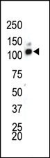
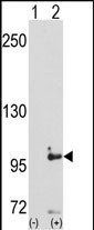
DDR1 Antibody (C-term)
Purified Rabbit Polyclonal Antibody (Pab)
- SPECIFICATION
- CITATIONS
- PROTOCOLS
- BACKGROUND

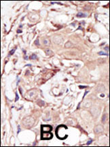
Application
| WB, IHC-P, E |
|---|---|
| Primary Accession | Q08345 |
| Reactivity | Human, Mouse |
| Host | Rabbit |
| Clonality | Polyclonal |
| Isotype | Rabbit IgG |
| Calculated MW | 101128 Da |
| Antigen Region | 873-904 aa |
| Gene ID | 780 |
|---|---|
| Other Names | Epithelial discoidin domain-containing receptor 1, Epithelial discoidin domain receptor 1, CD167 antigen-like family member A, Cell adhesion kinase, Discoidin receptor tyrosine kinase, HGK2, Mammary carcinoma kinase 10, MCK-10, Protein-tyrosine kinase 3A, Protein-tyrosine kinase RTK-6, TRK E, Tyrosine kinase DDR, Tyrosine-protein kinase CAK, CD167a, DDR1, CAK, EDDR1, NEP, NTRK4, PTK3A, RTK6, TRKE |
| Target/Specificity | This DDR1 antibody is generated from rabbits immunized with a KLH conjugated synthetic peptide between 873-904 amino acids from the C-terminal region of human DDR1. |
| Dilution | WB~~1:1000 IHC-P~~1:50~100 E~~Use at an assay dependent concentration. |
| Format | Purified polyclonal antibody supplied in PBS with 0.09% (W/V) sodium azide. This antibody is prepared by Saturated Ammonium Sulfate (SAS) precipitation followed by dialysis against PBS. |
| Storage | Maintain refrigerated at 2-8°C for up to 2 weeks. For long term storage store at -20°C in small aliquots to prevent freeze-thaw cycles. |
| Precautions | DDR1 Antibody (C-term) is for research use only and not for use in diagnostic or therapeutic procedures. |
| Name | DDR1 |
|---|---|
| Synonyms | CAK, EDDR1, NEP, NTRK4, PTK3A, RTK6, TRK |
| Function | Tyrosine kinase that functions as a cell surface receptor for fibrillar collagen and regulates cell attachment to the extracellular matrix, remodeling of the extracellular matrix, cell migration, differentiation, survival and cell proliferation. Collagen binding triggers a signaling pathway that involves SRC and leads to the activation of MAP kinases. Regulates remodeling of the extracellular matrix by up-regulation of the matrix metalloproteinases MMP2, MMP7 and MMP9, and thereby facilitates cell migration and wound healing. Required for normal blastocyst implantation during pregnancy, for normal mammary gland differentiation and normal lactation. Required for normal ear morphology and normal hearing (By similarity). Promotes smooth muscle cell migration, and thereby contributes to arterial wound healing. Also plays a role in tumor cell invasion. Phosphorylates PTPN11. |
| Cellular Location | [Isoform 1]: Cell membrane; Single-pass type I membrane protein [Isoform 3]: Secreted. |
| Tissue Location | Detected in T-47D, MDA-MB-175 and HBL-100 breast carcinoma cells, A-431 epidermoid carcinoma cells, SW48 and SNU-C2B colon carcinoma cells and Hs 294T melanoma cells (at protein level) Expressed at low levels in most adult tissues and is highest in the brain, lung, placenta and kidney. Lower levels of expression are detected in melanocytes, heart, liver, skeletal muscle and pancreas Abundant in breast carcinoma cell lines. In the colonic mucosa, expressed in epithelia but not in the connective tissue of the lamina propria. In the thyroid gland, expressed in the epithelium of the thyroid follicles. In pancreas, expressed in the islets of Langerhans cells, but not in the surrounding epithelial cells of the exocrine pancreas. In kidney, expressed in the epithelia of the distal tubules Not expressed in connective tissue, endothelial cells, adipose tissue, muscle cells or cells of hematopoietic origin |

Thousands of laboratories across the world have published research that depended on the performance of antibodies from Abcepta to advance their research. Check out links to articles that cite our products in major peer-reviewed journals, organized by research category.
info@abcepta.com, and receive a free "I Love Antibodies" mug.

Provided below are standard protocols that you may find useful for product applications.
Background
Receptor tyrosine kinases (RTKs) play a key role in the communication of cells with their microenvironment. These molecules are involved in the regulation of cell growth, differentiation and metabolism. MCK10 is a RTK that is widely expressed in normal and transformed epithelial cells and is activated by various types of collagen. This protein belongs to a subfamily of tyrosine kinase receptors with a homology region to the Dictyostelium discoideum protein discoidin I in their extracellular domain. Its autophosphorylation is achieved by all collagens so far tested (type I to type VI). In situ studies and Northern-blot analysis showed that expression of this encoded protein is restricted to epithelial cells, particularly in the kidney, lung, gastrointestinal tract, and brain. In addition, this protein is significantly over-expressed in several human tumors from breast, ovarian, esophageal, and pediatric brain. The gene is located on chromosome 6p21.3 in proximity to several HLA class I genes.
References
Vogel, W., et al., Mol. Cell 1(1):13-23 (1997).
Playford, M.P., et al., Genome Res. 6(7):620-627 (1996).
Perez, J.L., et al., Oncogene 12(7):1469-1477 (1996).
Valent, A., et al., Hum. Genet. 98(1):12-15 (1996).
Edelhoff, S., et al., Genomics 25(1):309-311 (1995).
If you have used an Abcepta product and would like to share how it has performed, please click on the "Submit Review" button and provide the requested information. Our staff will examine and post your review and contact you if needed.
If you have any additional inquiries please email technical services at tech@abcepta.com.
Ordering Information
Other Products
Shipping Information
