


 Foundational characteristics of cancer include proliferation, angiogenesis, migration, evasion of apoptosis, and cellular immortality. Find key markers for these cellular processes and antibodies to detect them.
 The SUMOplot™ Analysis Program predicts and scores sumoylation sites in your protein. SUMOylation is a post-translational modification involved in various cellular processes, such as nuclear-cytosolic transport, transcriptional regulation, apoptosis, protein stability, response to stress, and progression through the cell cycle.
 The Autophagy Receptor Motif Plotter predicts and scores autophagy receptor binding sites in your protein. Identifying proteins connected to this pathway is critical to understanding the role of autophagy in physiological as well as pathological processes such as development, differentiation, neurodegenerative diseases, stress, infection, and cancer.
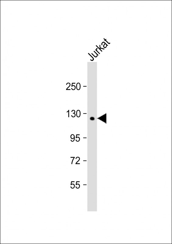
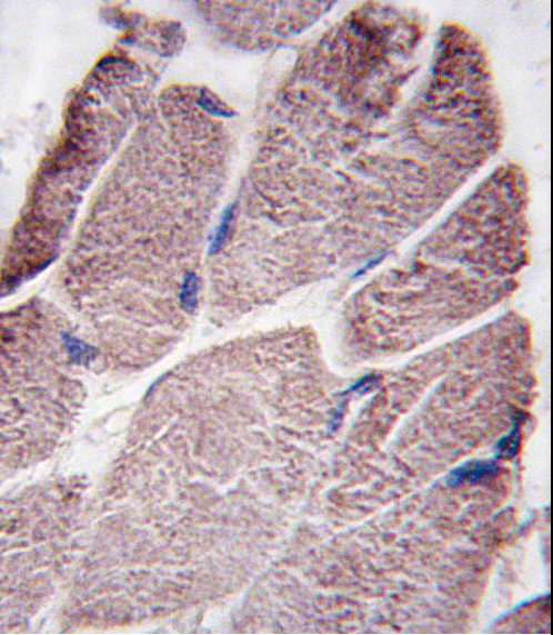
PTPD1 Antibody (Center)
Purified Rabbit Polyclonal Antibody (Pab)
- SPECIFICATION
- CITATIONS: 1
- PROTOCOLS
- BACKGROUND

Application
| IHC-P, WB, E |
|---|---|
| Primary Accession | Q16825 |
| Reactivity | Human |
| Host | Rabbit |
| Clonality | Polyclonal |
| Isotype | Rabbit IgG |
| Calculated MW | 133281 Da |
| Antigen Region | 751-780 aa |
| Gene ID | 11099 |
|---|---|
| Other Names | Tyrosine-protein phosphatase non-receptor type 21, Protein-tyrosine phosphatase D1, PTPN21, PTPD1 |
| Target/Specificity | This PTPD1 antibody is generated from rabbits immunized with a KLH conjugated synthetic peptide between 751-780 amino acids from the Central region of human PTPD1. |
| Dilution | IHC-P~~1:10~50 WB~~1:1000 E~~Use at an assay dependent concentration. |
| Format | Purified polyclonal antibody supplied in PBS with 0.09% (W/V) sodium azide. This antibody is prepared by Saturated Ammonium Sulfate (SAS) precipitation followed by dialysis against PBS. |
| Storage | Maintain refrigerated at 2-8°C for up to 2 weeks. For long term storage store at -20°C in small aliquots to prevent freeze-thaw cycles. |
| Precautions | PTPD1 Antibody (Center) is for research use only and not for use in diagnostic or therapeutic procedures. |
| Name | PTPN21 |
|---|---|
| Synonyms | PTPD1 |
| Cellular Location | Cytoplasm, cytoskeleton. |

Provided below are standard protocols that you may find useful for product applications.
Background
PTPD1 is a member of the protein tyrosine phosphatase (PTP) family. PTPs are known to be signaling molecules that regulate a variety of cellular processes including cell growth, differentiation, mitotic cycle, and oncogenic transformation. This PTP contains an N-terminal domain, similar to cytoskeletal- associated proteins including band 4.1, ezrin, merlin, and radixin. This PTP was shown to specially interact with BMX/ETK, a member of Tec tyrosine kinase family characterized by a multimodular structures including PH, SH3, and SH2 domains. The interaction of this PTP with BMX kinase was found to increase the activation of STAT3, but not STAT2 kinase. Studies of the similar gene in mice suggested the possible roles of this PTP in liver regeneration and spermatogenesis.
References
Jui, H.Y., et al., J. Biol. Chem. 275(52):41124-41132 (2000).
Tokuchi, H., et al., Int J Urol 6(11):572-577 (1999).
Higashitsuji, H., et al., Oncogene 10(2):407-414 (1995).
Moller, N.P., et al., Proc. Natl. Acad. Sci. U.S.A. 91(16):7477-7481 (1994).
If you have used an Abcepta product and would like to share how it has performed, please click on the "Submit Review" button and provide the requested information. Our staff will examine and post your review and contact you if needed.
If you have any additional inquiries please email technical services at tech@abcepta.com.
Ordering Information
Other Products
Shipping Information
