


 Foundational characteristics of cancer include proliferation, angiogenesis, migration, evasion of apoptosis, and cellular immortality. Find key markers for these cellular processes and antibodies to detect them.
 The SUMOplot™ Analysis Program predicts and scores sumoylation sites in your protein. SUMOylation is a post-translational modification involved in various cellular processes, such as nuclear-cytosolic transport, transcriptional regulation, apoptosis, protein stability, response to stress, and progression through the cell cycle.
 The Autophagy Receptor Motif Plotter predicts and scores autophagy receptor binding sites in your protein. Identifying proteins connected to this pathway is critical to understanding the role of autophagy in physiological as well as pathological processes such as development, differentiation, neurodegenerative diseases, stress, infection, and cancer.
CAT Antibody (Center)
Affinity Purified Rabbit Polyclonal Antibody (Pab)
- SPECIFICATION
- CITATIONS: 2
- PROTOCOLS
- BACKGROUND

Application
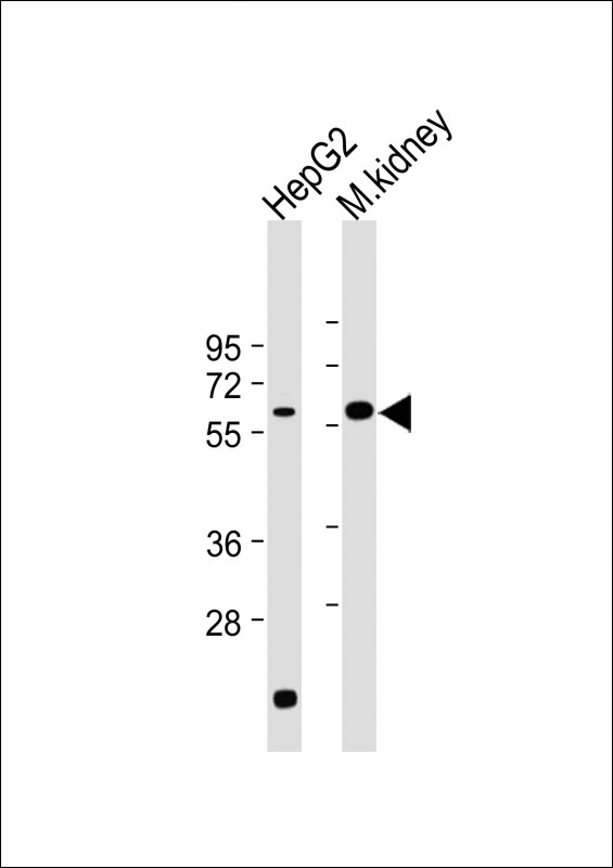
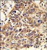
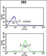
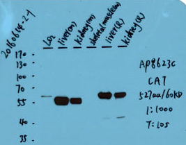
| FC, IHC-P, WB, E |
|---|---|
| Primary Accession | P04040 |
| Other Accession | P04762, O62839, P24270, Q9PT92, P00432 |
| Reactivity | Human, Mouse |
| Predicted | Bovine, Zebrafish, Pig, Rat |
| Host | Rabbit |
| Clonality | Polyclonal |
| Isotype | Rabbit IgG |
| Calculated MW | 59756 Da |
| Antigen Region | 152-180 aa |
| Gene ID | 847 |
|---|---|
| Other Names | Catalase, CAT |
| Target/Specificity | This CAT antibody is generated from rabbits immunized with a KLH conjugated synthetic peptide between 152-180 amino acids from the Central region of human CAT. |
| Dilution | FC~~1:10~50 IHC-P~~1:50~100 WB~~1:2000 E~~Use at an assay dependent concentration. |
| Format | Purified polyclonal antibody supplied in PBS with 0.09% (W/V) sodium azide. This antibody is purified through a protein A column, followed by peptide affinity purification. |
| Storage | Maintain refrigerated at 2-8°C for up to 2 weeks. For long term storage store at -20°C in small aliquots to prevent freeze-thaw cycles. |
| Precautions | CAT Antibody (Center) is for research use only and not for use in diagnostic or therapeutic procedures. |
| Name | CAT |
|---|---|
| Function | Catalyzes the degradation of hydrogen peroxide (H(2)O(2)) generated by peroxisomal oxidases to water and oxygen, thereby protecting cells from the toxic effects of hydrogen peroxide (PubMed:7882369). Promotes growth of cells including T-cells, B-cells, myeloid leukemia cells, melanoma cells, mastocytoma cells and normal and transformed fibroblast cells (PubMed:7882369). |
| Cellular Location | Peroxisome matrix |

Provided below are standard protocols that you may find useful for product applications.
Background
CAT occurs in almost all aerobically respiring organisms and serves to protect cells from the toxic effects of hydrogen peroxide. It promotes growth of cells including T-cells, B-cells, myeloid leukemia cells, melanoma cells, mastocytoma cells and normal and transformed fibroblast cells.
References
Oh,J.H., et.al., Mamm. Genome 16 (12), 942-954 (2005)
If you have used an Abcepta product and would like to share how it has performed, please click on the "Submit Review" button and provide the requested information. Our staff will examine and post your review and contact you if needed.
If you have any additional inquiries please email technical services at tech@abcepta.com.
Anonymous
2016-08-24 01:15:32
1
2
3
4
5
|
Species tested
Human,Mouse,Rat
Application tested
WB
Brief protocol
1. Block with 3% skim milk for 1 hour at room temperature.
2. Incubate overnight with Abgent primary antibody 1:1000 in 3% skim milk at 4℃
3. Wash 5*5 min with TBST.
4. Incubate with HRP-conjugated secondary antibody 1:5000 in 3% skim milk for 1 hour at room temperature.
5. Wash 5*5 min with TBST.
6. Incubate with ECL substrates and expose |
 |
Ordering Information
Other Products
Shipping Information
