


 Foundational characteristics of cancer include proliferation, angiogenesis, migration, evasion of apoptosis, and cellular immortality. Find key markers for these cellular processes and antibodies to detect them.
 The SUMOplot™ Analysis Program predicts and scores sumoylation sites in your protein. SUMOylation is a post-translational modification involved in various cellular processes, such as nuclear-cytosolic transport, transcriptional regulation, apoptosis, protein stability, response to stress, and progression through the cell cycle.
 The Autophagy Receptor Motif Plotter predicts and scores autophagy receptor binding sites in your protein. Identifying proteins connected to this pathway is critical to understanding the role of autophagy in physiological as well as pathological processes such as development, differentiation, neurodegenerative diseases, stress, infection, and cancer.
KLC1 Antibody (Center)
Affinity Purified Rabbit Polyclonal Antibody (Pab)
- SPECIFICATION
- CITATIONS: 1
- PROTOCOLS
- BACKGROUND

Application
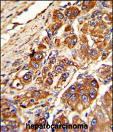
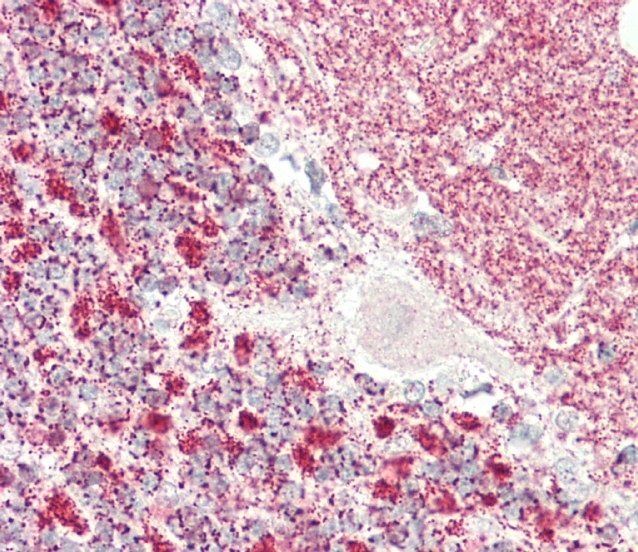
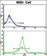
| WB, FC, IHC-P, E |
|---|---|
| Primary Accession | Q07866 |
| Other Accession | P37285 |
| Reactivity | Human |
| Predicted | Rat |
| Host | Rabbit |
| Clonality | Polyclonal |
| Isotype | Rabbit IgG |
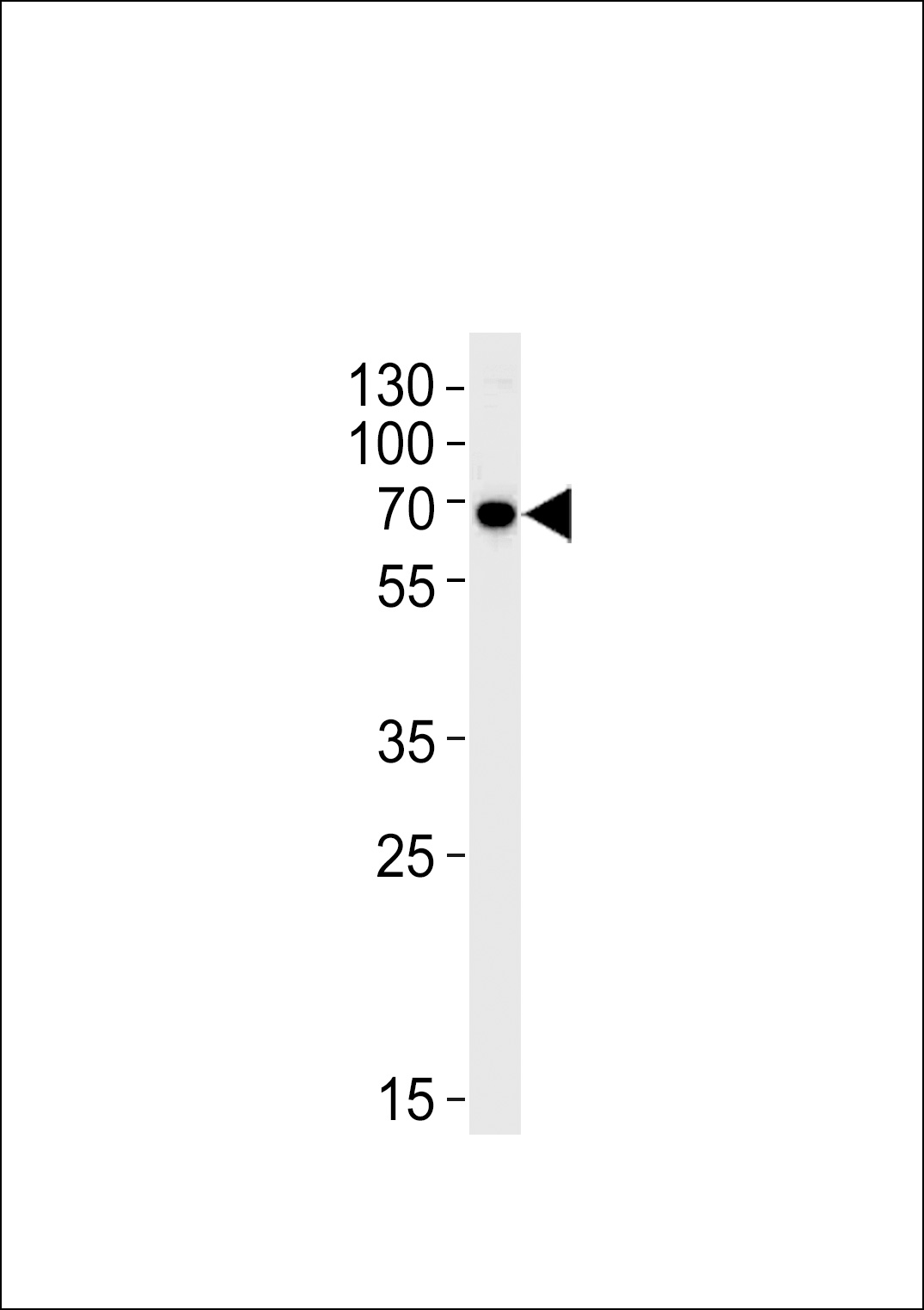
| Calculated MW | 65310 Da |
| Antigen Region | 389-415 aa |
| Gene ID | 3831 |
|---|---|
| Other Names | Kinesin light chain 1, KLC 1, KLC1, KLC, KNS2 |
| Target/Specificity | This KLC1 antibody is generated from rabbits immunized with a KLH conjugated synthetic peptide between 389-415 amino acids from the Central region of human KLC1. |
| Dilution | WB~~1:1000 FC~~1:10~50 IHC-P~~1:100 E~~Use at an assay dependent concentration. |
| Format | Purified polyclonal antibody supplied in PBS with 0.09% (W/V) sodium azide. This antibody is purified through a protein A column, followed by peptide affinity purification. |
| Storage | Maintain refrigerated at 2-8°C for up to 2 weeks. For long term storage store at -20°C in small aliquots to prevent freeze-thaw cycles. |
| Precautions | KLC1 Antibody (Center) is for research use only and not for use in diagnostic or therapeutic procedures. |
| Name | KLC1 |
|---|---|
| Synonyms | KLC, KNS2 |
| Function | Kinesin is a microtubule-associated force-producing protein that may play a role in organelle transport (PubMed:21385839). The light chain may function in coupling of cargo to the heavy chain or in the modulation of its ATPase activity (By similarity). |
| Cellular Location | Cell projection, growth cone {ECO:0000250|UniProtKB:P37285}. Cytoplasmic vesicle. Cytoplasm, cytoskeleton |
| Tissue Location | Found in a variety of tissues. Mostly abundant in brain and spine. |

Provided below are standard protocols that you may find useful for product applications.
Background
Kinesin is a microtubule-associated force-producing protein that may play a role in organelle transport. The light chain may function in coupling of cargo to the heavy chain or in the modulation of its ATPase activity.
References
Chernajovsky,Y., et.al., DNA Cell Biol. 15 (11), 965-974 (1996) Gyoeva,F.K., et.al., J. Cell. Sci. 113 (PT 11), 2047-2054 (2000)
If you have used an Abcepta product and would like to share how it has performed, please click on the "Submit Review" button and provide the requested information. Our staff will examine and post your review and contact you if needed.
If you have any additional inquiries please email technical services at tech@abcepta.com.
Ordering Information
Other Products
Shipping Information
