


 Foundational characteristics of cancer include proliferation, angiogenesis, migration, evasion of apoptosis, and cellular immortality. Find key markers for these cellular processes and antibodies to detect them.
 The SUMOplot™ Analysis Program predicts and scores sumoylation sites in your protein. SUMOylation is a post-translational modification involved in various cellular processes, such as nuclear-cytosolic transport, transcriptional regulation, apoptosis, protein stability, response to stress, and progression through the cell cycle.
 The Autophagy Receptor Motif Plotter predicts and scores autophagy receptor binding sites in your protein. Identifying proteins connected to this pathway is critical to understanding the role of autophagy in physiological as well as pathological processes such as development, differentiation, neurodegenerative diseases, stress, infection, and cancer.
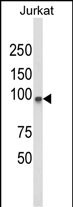
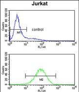
PLCL1 Antibody (N-term)
Affinity Purified Rabbit Polyclonal Antibody (Pab)
- SPECIFICATION
- CITATIONS
- PROTOCOLS
- BACKGROUND

Application
| FC, WB, E |
|---|---|
| Primary Accession | Q15111 |
| Other Accession | Q62688, Q3USB7 |
| Reactivity | Human |
| Predicted | Mouse, Rat |
| Host | Rabbit |
| Clonality | Polyclonal |
| Isotype | Rabbit IgG |
| Calculated MW | 122728 Da |
| Antigen Region | 149-176 aa |
| Gene ID | 5334 |
|---|---|
| Other Names | Inactive phospholipase C-like protein 1, PLC-L1, Phospholipase C-deleted in lung carcinoma, Phospholipase C-related but catalytically inactive protein, PRIP, PLCL1 |
| Target/Specificity | This PLCL1 antibody is generated from rabbits immunized with a KLH conjugated synthetic peptide between 149-176 amino acids from the N-terminal region of human PLCL1. |
| Dilution | FC~~1:10~50 WB~~1:1000 E~~Use at an assay dependent concentration. |
| Format | Purified polyclonal antibody supplied in PBS with 0.09% (W/V) sodium azide. This antibody is purified through a protein A column, followed by peptide affinity purification. |
| Storage | Maintain refrigerated at 2-8°C for up to 2 weeks. For long term storage store at -20°C in small aliquots to prevent freeze-thaw cycles. |
| Precautions | PLCL1 Antibody (N-term) is for research use only and not for use in diagnostic or therapeutic procedures. |
| Name | PLCL1 |
|---|---|
| Function | Involved in an inositol phospholipid-based intracellular signaling cascade. Shows no PLC activity to phosphatidylinositol 4,5- bisphosphate and phosphatidylinositol. Component in the phospho- dependent endocytosis process of GABA A receptor (By similarity). Regulates the turnover of receptors and thus contributes to the maintenance of GABA-mediated synaptic inhibition. Its aberrant expression could contribute to the genesis and progression of lung carcinoma. Acts as an inhibitor of PPP1C. |
| Cellular Location | Cytoplasm. |
| Tissue Location | Expressed in a variety of fetal and adult organs including brain, lung and kidney. Its expression was greatly reduced in small and non-small cell lung carcinoma. Isoform 1 is predominantly expressed in brain. |

Thousands of laboratories across the world have published research that depended on the performance of antibodies from Abcepta to advance their research. Check out links to articles that cite our products in major peer-reviewed journals, organized by research category.
info@abcepta.com, and receive a free "I Love Antibodies" mug.

Provided below are standard protocols that you may find useful for product applications.
Background
PLCL1 is an inositol phospholipid-based intracellular signaling cascade. Shows no PLC activity to phosphatidylinositol 4,5-bisphosphate and sphatidylinositol. Component in the phospho-dependent endocytosis process of GABA A receptor (By similarity). Regulates the turnover of receptors and thus contributes to the maintenance of GABA-mediated synaptic inhibition. Its aberrant expression could contribute to the genesis and progression of lung carcinoma. Acts as a nhibitor of PPP1C.
References
Liu,Y.Z., et.al., PLoS ONE 3 (9), E3160 (2008)
If you have used an Abcepta product and would like to share how it has performed, please click on the "Submit Review" button and provide the requested information. Our staff will examine and post your review and contact you if needed.
If you have any additional inquiries please email technical services at tech@abcepta.com.
Ordering Information
Shipping Information
