


 Foundational characteristics of cancer include proliferation, angiogenesis, migration, evasion of apoptosis, and cellular immortality. Find key markers for these cellular processes and antibodies to detect them.
 The SUMOplot™ Analysis Program predicts and scores sumoylation sites in your protein. SUMOylation is a post-translational modification involved in various cellular processes, such as nuclear-cytosolic transport, transcriptional regulation, apoptosis, protein stability, response to stress, and progression through the cell cycle.
 The Autophagy Receptor Motif Plotter predicts and scores autophagy receptor binding sites in your protein. Identifying proteins connected to this pathway is critical to understanding the role of autophagy in physiological as well as pathological processes such as development, differentiation, neurodegenerative diseases, stress, infection, and cancer.
TBB1 Antibody
Affinity Purified Rabbit Polyclonal Antibody (Pab)
- SPECIFICATION
- CITATIONS
- PROTOCOLS
- BACKGROUND

Application
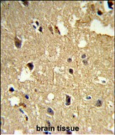
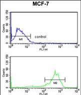
| FC, IHC-P, WB, E |
|---|---|
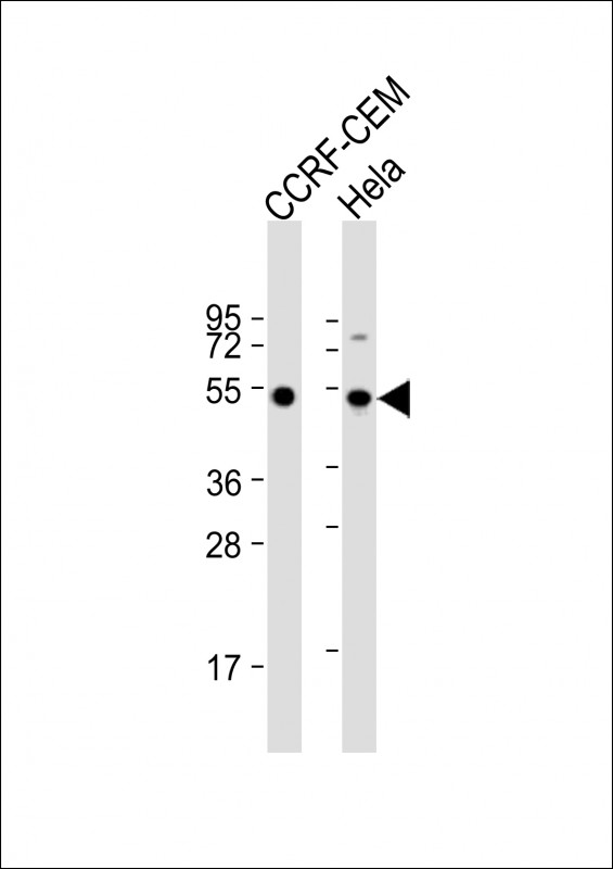
| Primary Accession | Q9H4B7 |
| Reactivity | Human |
| Host | Rabbit |
| Clonality | Polyclonal |
| Isotype | Rabbit IgG |
| Calculated MW | 50327 Da |
| Gene ID | 81027 |
|---|---|
| Other Names | Tubulin beta-1 chain, TUBB1 |
| Target/Specificity | This TBB1 antibody is generated from rabbits immunized with human TBB1 recombinant protein. |
| Dilution | FC~~1:10~50 IHC-P~~1:50~100 WB~~1:2000 E~~Use at an assay dependent concentration. |
| Format | Purified polyclonal antibody supplied in PBS with 0.09% (W/V) sodium azide. This antibody is purified through a protein A column, followed by peptide affinity purification. |
| Storage | Maintain refrigerated at 2-8°C for up to 2 weeks. For long term storage store at -20°C in small aliquots to prevent freeze-thaw cycles. |
| Precautions | TBB1 Antibody is for research use only and not for use in diagnostic or therapeutic procedures. |
| Name | TUBB1 |
|---|---|
| Function | Tubulin is the major constituent of microtubules, a cylinder consisting of laterally associated linear protofilaments composed of alpha- and beta-tubulin heterodimers. Microtubules grow by the addition of GTP-tubulin dimers to the microtubule end, where a stabilizing cap forms. Below the cap, tubulin dimers are in GDP-bound state, owing to GTPase activity of alpha-tubulin. |
| Cellular Location | Cytoplasm, cytoskeleton |
| Tissue Location | Hematopoietic cell-specific. Major isotype in leukocytes, where it represents 50% of all beta-tubulins |

Thousands of laboratories across the world have published research that depended on the performance of antibodies from Abcepta to advance their research. Check out links to articles that cite our products in major peer-reviewed journals, organized by research category.
info@abcepta.com, and receive a free "I Love Antibodies" mug.

Provided below are standard protocols that you may find useful for product applications.
Background
The tubulin family of globular proteins has several members, the most common of which are a-tubulin and ?tubulin; proteins which make up microtubules of the cytoskeltons of probably all eukaryotic cells. Except in the simplest eukaryotes, tubulin (100 kDa) exists in all cells as a heterodimer of two similar but non-identical polypeptides (55 kDa each), designated alpha and beta. Within either family of alpha/beta tubulin heterodimers, individual subunits diverge from each other (both within and across species) at less than 10% of the amino acid positions. The most extreme diversity is localized to the carboxyl-terminal 15 residues. Delta (d) and epsilon (e) tubulin have been found to localize at centrioles and may play a role in forming the mitotic spindle during mitosis, though neither is as well-studied as the a- and ? forms.
References
Rogowski K., et.al., Cell 137:1076-1087(2009).
If you have used an Abcepta product and would like to share how it has performed, please click on the "Submit Review" button and provide the requested information. Our staff will examine and post your review and contact you if needed.
If you have any additional inquiries please email technical services at tech@abcepta.com.
Ordering Information
Other Products
Shipping Information
