


 Foundational characteristics of cancer include proliferation, angiogenesis, migration, evasion of apoptosis, and cellular immortality. Find key markers for these cellular processes and antibodies to detect them.
 The SUMOplot™ Analysis Program predicts and scores sumoylation sites in your protein. SUMOylation is a post-translational modification involved in various cellular processes, such as nuclear-cytosolic transport, transcriptional regulation, apoptosis, protein stability, response to stress, and progression through the cell cycle.
 The Autophagy Receptor Motif Plotter predicts and scores autophagy receptor binding sites in your protein. Identifying proteins connected to this pathway is critical to understanding the role of autophagy in physiological as well as pathological processes such as development, differentiation, neurodegenerative diseases, stress, infection, and cancer.
LYVE1 Antibody
Rabbit mAb
- SPECIFICATION
- CITATIONS
- PROTOCOLS
- BACKGROUND

Application
| WB |
|---|---|
| Primary Accession | Q9Y5Y7 |
| Reactivity | Rat |
| Clonality | Monoclonal |
| Other Names | CRSBP 1; CRSBP-1; CRSBP1; hyaluronic acid receptor; |
| Isotype | Rabbit IgG |
| Host | Rabbit |
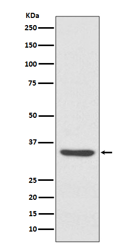
| Calculated MW | 35213 Da |
| Dilution | WB 1:500~1:2000 |
|---|---|
| Purification | Affinity-chromatography |
| Immunogen | A synthesized peptide derived from human LYVE1 |
| Description | Plays a role in autocrine regulation of cell growth mediated by growth regulators containing cell surface retention sequence binding (CRS). May act as a hyaluronan (HA) transporter, either mediating its uptake for catabolism within lymphatic endothelial cells themselves, or its transport into the lumen of afferent lymphatic vessels for subsequent re-uptake and degradation in lymph nodes. |
| Storage Condition and Buffer | Rabbit IgG in phosphate buffered saline , pH 7.4, 150mM NaCl, 0.02% sodium azide and 50% glycerol. Store at +4°C short term. Store at -20°C long term. Avoid freeze / thaw cycle. |
| Name | LYVE1 |
|---|---|
| Synonyms | CRSBP1, HAR, XLKD1 |
| Function | Ligand-specific transporter trafficking between intracellular organelles (TGN) and the plasma membrane. Plays a role in autocrine regulation of cell growth mediated by growth regulators containing cell surface retention sequence binding (CRS). May act as a hyaluronan (HA) transporter, either mediating its uptake for catabolism within lymphatic endothelial cells themselves, or its transport into the lumen of afferent lymphatic vessels for subsequent re-uptake and degradation in lymph nodes (PubMed:10037799). Binds to pericelluar hyaluronan matrices deposited on the surface of leukocytes and facilitates cell adhesion and migration through lymphatic endothelium (PubMed:26823460). |
| Cellular Location | Cell membrane; Single-pass type I membrane protein. Note=Localized to the plasma membrane and in vesicles near extranuclear membranes which may represent trans- Golgi network (TGN) and endosomes/prelysosomeal compartments. Undergoes ligand-dependent internalization and recycling at the cell surface Localizes at cell-cell junctions |
| Tissue Location | Mainly expressed in endothelial cells lining lymphatic vessels. |
Research Areas
Citations (0)

Thousands of laboratories across the world have published research that depended on the performance of antibodies from Abcepta to advance their research. Check out links to articles that cite our products in major peer-reviewed journals, organized by research category.
Submit your citation using an Abcepta antibody to
info@abcepta.com, and receive a free "I Love Antibodies" mug.
info@abcepta.com, and receive a free "I Love Antibodies" mug.

Application Protocols
Provided below are standard protocols that you may find useful for product applications.
Abcepta welcomes feedback from its customers.
If you have used an Abcepta product and would like to share how it has performed, please click on the "Submit Review" button and provide the requested information. Our staff will examine and post your review and contact you if needed.
If you have any additional inquiries please email technical services at tech@abcepta.com.
$ 195.00
$ 385.00
Cat# AP91068
Ordering Information
United States
AlbaniaAustraliaAustriaBelgiumBosnia & HerzegovinaBrazilBulgariaCanadaCentral AmericaChinaCroatiaCyprusCzech RepublicDenmarkEstoniaFinlandFranceGermanyGreeceHong KongHungaryIcelandIndiaIndonesiaIrelandIsraelItalyJapanLatviaLithuaniaLuxembourgMacedoniaMalaysiaMaltaMexicoNetherlandsNew ZealandNorwayPakistanPolandPortugalRomaniaSerbiaSingaporeSlovakiaSloveniaSouth AfricaSouth KoreaSpainSwedenSwitzerlandTaiwanTurkeyUnited KingdomUnited StatesVietnamWorldwideOthers
USA Headquarters
(888) 735-7227 / (858) 622-0099 or (858) 875-1900
Other Products
Shipping Information
Domestic orders (in stock items)
Shipped out the same day. Orders placed after 1 PM (PST) will ship out the next business day.
International orders
Contact your local distributors
