


 Foundational characteristics of cancer include proliferation, angiogenesis, migration, evasion of apoptosis, and cellular immortality. Find key markers for these cellular processes and antibodies to detect them.
 The SUMOplot™ Analysis Program predicts and scores sumoylation sites in your protein. SUMOylation is a post-translational modification involved in various cellular processes, such as nuclear-cytosolic transport, transcriptional regulation, apoptosis, protein stability, response to stress, and progression through the cell cycle.
 The Autophagy Receptor Motif Plotter predicts and scores autophagy receptor binding sites in your protein. Identifying proteins connected to this pathway is critical to understanding the role of autophagy in physiological as well as pathological processes such as development, differentiation, neurodegenerative diseases, stress, infection, and cancer.
ILF2 Antibody
Rabbit mAb
- SPECIFICATION
- CITATIONS
- PROTOCOLS
- BACKGROUND

Application
| WB, IHC, FC, IP |
|---|---|
| Primary Accession | Q12905 |
| Reactivity | Rat |
| Clonality | Monoclonal |
| Other Names | ILF 2; MGC8391; NF45; |
| Isotype | Rabbit IgG |
| Host | Rabbit |
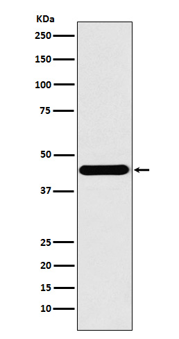
| Calculated MW | 43062 Da |
| Dilution | WB 1:500~1:2000 IHC 1:50~1:200 IP 1:50 FC 1:100 |
|---|---|
| Purification | Affinity-chromatography |
| Immunogen | A synthesized peptide derived from human ILF2 |
| Description | Appears to function predominantly as a heterodimeric complex with ILF3. This complex may regulate transcription of the IL2 gene during T-cell activation. It can also promote the formation of stable DNA-dependent protein kinase holoenzyme complexes on DNA. |
| Storage Condition and Buffer | Rabbit IgG in phosphate buffered saline , pH 7.4, 150mM NaCl, 0.02% sodium azide and 50% glycerol. Store at +4°C short term. Store at -20°C long term. Avoid freeze / thaw cycle. |
| Name | ILF2 |
|---|---|
| Synonyms | NF45 |
| Function | Chromatin-interacting protein that forms a stable heterodimer with interleukin enhancer-binding factor 3/ILF3 and plays a role in several biological processes including transcription, innate immunity or cell growth (PubMed:18458058, PubMed:31212927). Essential for the efficient reshuttling of ILF3 (isoform 1 and isoform 2) into the nucleus. Together with ILF3, forms an RNA-binding complex that is required for mitotic progression and cytokinesis by regulating the expression of a cluster of mitotic genes. Mechanistically, competes with STAU1/STAU2-mediated mRNA decay (PubMed:32433969). Also plays a role in the inhibition of various viruses including Japanese encephalitis virus or enterovirus 71. |
| Cellular Location | Nucleus, nucleolus. Cytoplasm. Nucleus. Note=Localized in cytoplasmic mRNP granules containing untranslated mRNAs |
Citations (0)

Thousands of laboratories across the world have published research that depended on the performance of antibodies from Abcepta to advance their research. Check out links to articles that cite our products in major peer-reviewed journals, organized by research category.
Submit your citation using an Abcepta antibody to
info@abcepta.com, and receive a free "I Love Antibodies" mug.
info@abcepta.com, and receive a free "I Love Antibodies" mug.

Application Protocols
Provided below are standard protocols that you may find useful for product applications.
Abcepta welcomes feedback from its customers.
If you have used an Abcepta product and would like to share how it has performed, please click on the "Submit Review" button and provide the requested information. Our staff will examine and post your review and contact you if needed.
If you have any additional inquiries please email technical services at tech@abcepta.com.
$ 195.00
$ 385.00
Cat# AP91698
Ordering Information
United States
AlbaniaAustraliaAustriaBelgiumBosnia & HerzegovinaBrazilBulgariaCanadaCentral AmericaChinaCroatiaCyprusCzech RepublicDenmarkEstoniaFinlandFranceGermanyGreeceHong KongHungaryIcelandIndiaIndonesiaIrelandIsraelItalyJapanLatviaLithuaniaLuxembourgMacedoniaMalaysiaMaltaMexicoNetherlandsNew ZealandNorwayPakistanPolandPortugalRomaniaSerbiaSingaporeSlovakiaSloveniaSouth AfricaSouth KoreaSpainSwedenSwitzerlandTaiwanTurkeyUnited KingdomUnited StatesVietnamWorldwideOthers
USA Headquarters
(888) 735-7227 / (858) 622-0099 or (858) 875-1900
Other Products
Shipping Information
Domestic orders (in stock items)
Shipped out the same day. Orders placed after 1 PM (PST) will ship out the next business day.
International orders
Contact your local distributors
