


 Foundational characteristics of cancer include proliferation, angiogenesis, migration, evasion of apoptosis, and cellular immortality. Find key markers for these cellular processes and antibodies to detect them.
 The SUMOplot™ Analysis Program predicts and scores sumoylation sites in your protein. SUMOylation is a post-translational modification involved in various cellular processes, such as nuclear-cytosolic transport, transcriptional regulation, apoptosis, protein stability, response to stress, and progression through the cell cycle.
 The Autophagy Receptor Motif Plotter predicts and scores autophagy receptor binding sites in your protein. Identifying proteins connected to this pathway is critical to understanding the role of autophagy in physiological as well as pathological processes such as development, differentiation, neurodegenerative diseases, stress, infection, and cancer.
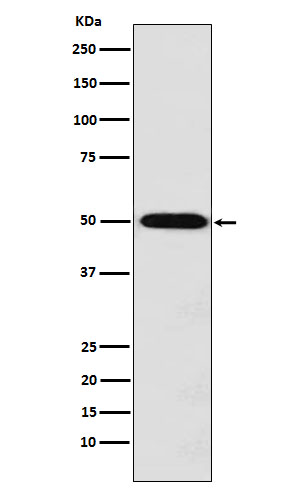
MCT1 Antibody
Rabbit mAb
- SPECIFICATION
- CITATIONS
- PROTOCOLS
- BACKGROUND

Application
| WB |
|---|---|
| Primary Accession | P53985 |
| Clonality | Monoclonal |
| Other Names | HHF7; MCT 1; MCT; Slc16a1; |
| Isotype | Rabbit IgG |
| Host | Rabbit |
| Calculated MW | 53944 Da |
| Dilution | WB 1:500~1:2000 |
|---|---|
| Purification | Affinity-chromatography |
| Immunogen | A synthesized peptide derived from human Monocarboxylic acid transporter 1 |
| Description | Proton-linked monocarboxylate transporter. Catalyzes the rapid transport across the plasma membrane of many monocarboxylates such as lactate, pyruvate, branched-chain oxo acids derived from leucine, valine and isoleucine, and the ketone bodies acetoacetate, beta-hydroxybutyrate and acetate. |
| Storage Condition and Buffer | Rabbit IgG in phosphate buffered saline , pH 7.4, 150mM NaCl, 0.02% sodium azide and 50% glycerol. Store at +4°C short term. Store at -20°C long term. Avoid freeze / thaw cycle. |
| Name | SLC16A1 (HGNC:10922) |
|---|---|
| Synonyms | MCT1 |
| Function | Bidirectional proton-coupled monocarboxylate transporter (PubMed:12946269, PubMed:32946811, PubMed:33333023). Catalyzes the rapid transport across the plasma membrane of many monocarboxylates such as lactate, pyruvate, acetate and the ketone bodies acetoacetate and beta-hydroxybutyrate, and thus contributes to the maintenance of intracellular pH (PubMed:12946269, PubMed:33333023). The transport direction is determined by the proton motive force and the concentration gradient of the substrate monocarboxylate. MCT1 is a major lactate exporter (By similarity). Plays a role in cellular responses to a high-fat diet by modulating the cellular levels of lactate and pyruvate that contribute to the regulation of central metabolic pathways and insulin secretion, with concomitant effects on plasma insulin levels and blood glucose homeostasis (By similarity). Facilitates the protonated monocarboxylate form of succinate export, that its transient protonation upon muscle cell acidification in exercising muscle and ischemic heart (PubMed:32946811). Functions via alternate outward- and inward-open conformation states. Protonation and deprotonation of 309-Asp is essential for the conformational transition (PubMed:33333023). |
| Cellular Location | Cell membrane; Multi-pass membrane protein. Basolateral cell membrane {ECO:0000250|UniProtKB:P53987}; Multi-pass membrane protein. Apical cell membrane; Multi-pass membrane protein {ECO:0000250|UniProtKB:P53987}. Note=Expression at the cell surface requires the ancillary proteins BSG and EMB. Binds preferentially to BSG. |
| Tissue Location | Widely expressed (PubMed:12115955, PubMed:15505343, PubMed:15901598). Detected in heart and in blood lymphocytes and monocytes (at protein level) (PubMed:15505343) |
Research Areas
Citations (0)

Thousands of laboratories across the world have published research that depended on the performance of antibodies from Abcepta to advance their research. Check out links to articles that cite our products in major peer-reviewed journals, organized by research category.
Submit your citation using an Abcepta antibody to
info@abcepta.com, and receive a free "I Love Antibodies" mug.
info@abcepta.com, and receive a free "I Love Antibodies" mug.

Application Protocols
Provided below are standard protocols that you may find useful for product applications.
Abcepta welcomes feedback from its customers.
If you have used an Abcepta product and would like to share how it has performed, please click on the "Submit Review" button and provide the requested information. Our staff will examine and post your review and contact you if needed.
If you have any additional inquiries please email technical services at tech@abcepta.com.
$ 195.00
$ 385.00
Cat# AP92044
Ordering Information
United States
AlbaniaAustraliaAustriaBelgiumBosnia & HerzegovinaBrazilBulgariaCanadaCentral AmericaChinaCroatiaCyprusCzech RepublicDenmarkEstoniaFinlandFranceGermanyGreeceHong KongHungaryIcelandIndiaIndonesiaIrelandIsraelItalyJapanLatviaLithuaniaLuxembourgMacedoniaMalaysiaMaltaMexicoNetherlandsNew ZealandNorwayPakistanPolandPortugalRomaniaSerbiaSingaporeSlovakiaSloveniaSouth AfricaSouth KoreaSpainSwedenSwitzerlandTaiwanTurkeyUnited KingdomUnited StatesVietnamWorldwideOthers
USA Headquarters
(888) 735-7227 / (858) 622-0099 or (858) 875-1900
Other Products
Shipping Information
Domestic orders (in stock items)
Shipped out the same day. Orders placed after 1 PM (PST) will ship out the next business day.
International orders
Contact your local distributors
