


 Foundational characteristics of cancer include proliferation, angiogenesis, migration, evasion of apoptosis, and cellular immortality. Find key markers for these cellular processes and antibodies to detect them.
 The SUMOplot™ Analysis Program predicts and scores sumoylation sites in your protein. SUMOylation is a post-translational modification involved in various cellular processes, such as nuclear-cytosolic transport, transcriptional regulation, apoptosis, protein stability, response to stress, and progression through the cell cycle.
 The Autophagy Receptor Motif Plotter predicts and scores autophagy receptor binding sites in your protein. Identifying proteins connected to this pathway is critical to understanding the role of autophagy in physiological as well as pathological processes such as development, differentiation, neurodegenerative diseases, stress, infection, and cancer.
EMC4 Antibody
Rabbit mAb
- SPECIFICATION
- CITATIONS
- PROTOCOLS
- BACKGROUND

Application
| WB, IHC, IP |
|---|---|
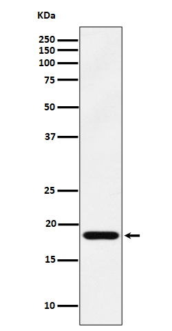
| Primary Accession | Q5J8M3 |
| Reactivity | Rat |
| Clonality | Monoclonal |
| Other Names | EMC4; PIG17; tmem85; Transmembrane protein 85; |
| Isotype | Rabbit IgG |
| Host | Rabbit |
| Calculated MW | 20087 Da |
| Dilution | WB 1:500~1:2000 IHC 1:50~1:200 IP 1:50 |
|---|---|
| Purification | Affinity-chromatography |
| Immunogen | A synthesized peptide derived from human EMC4 |
| Description | May mediate anti-apoptotic activity. |
| Storage Condition and Buffer | Rabbit IgG in phosphate buffered saline , pH 7.4, 150mM NaCl, 0.02% sodium azide and 50% glycerol. Store at +4°C short term. Store at -20°C long term. Avoid freeze / thaw cycle. |
| Name | EMC4 |
|---|---|
| Synonyms | TMEM85 |
| Function | Part of the endoplasmic reticulum membrane protein complex (EMC) that enables the energy-independent insertion into endoplasmic reticulum membranes of newly synthesized membrane proteins (PubMed:29242231, PubMed:29809151, PubMed:30415835, PubMed:32439656, PubMed:32459176). Preferentially accommodates proteins with transmembrane domains that are weakly hydrophobic or contain destabilizing features such as charged and aromatic residues (PubMed:29242231, PubMed:29809151, PubMed:30415835). Involved in the cotranslational insertion of multi-pass membrane proteins in which stop-transfer membrane-anchor sequences become ER membrane spanning helices (PubMed:29809151, PubMed:30415835). It is also required for the post-translational insertion of tail-anchored/TA proteins in endoplasmic reticulum membranes (PubMed:29242231, PubMed:29809151). By mediating the proper cotranslational insertion of N-terminal transmembrane domains in an N-exo topology, with translocated N- terminus in the lumen of the ER, controls the topology of multi-pass membrane proteins like the G protein-coupled receptors (PubMed:30415835). By regulating the insertion of various proteins in membranes, it is indirectly involved in many cellular processes (Probable). |
| Cellular Location | Endoplasmic reticulum membrane; Multi-pass membrane protein. Note=Could also be a single-pass transmembrane protein with cytosolic N-terminus and lumenal C-terminus. |
| Tissue Location | Isoform 1 is expressed in brain and heart. Isoform 2 is expressed in heart. |
Research Areas
Citations (0)

Thousands of laboratories across the world have published research that depended on the performance of antibodies from Abcepta to advance their research. Check out links to articles that cite our products in major peer-reviewed journals, organized by research category.
Submit your citation using an Abcepta antibody to
info@abcepta.com, and receive a free "I Love Antibodies" mug.
info@abcepta.com, and receive a free "I Love Antibodies" mug.

Application Protocols
Provided below are standard protocols that you may find useful for product applications.
Abcepta welcomes feedback from its customers.
If you have used an Abcepta product and would like to share how it has performed, please click on the "Submit Review" button and provide the requested information. Our staff will examine and post your review and contact you if needed.
If you have any additional inquiries please email technical services at tech@abcepta.com.
$ 195.00
$ 385.00
Cat# AP93036
Ordering Information
United States
AlbaniaAustraliaAustriaBelgiumBosnia & HerzegovinaBrazilBulgariaCanadaCentral AmericaChinaCroatiaCyprusCzech RepublicDenmarkEstoniaFinlandFranceGermanyGreeceHong KongHungaryIcelandIndiaIndonesiaIrelandIsraelItalyJapanLatviaLithuaniaLuxembourgMacedoniaMalaysiaMaltaMexicoNetherlandsNew ZealandNorwayPakistanPolandPortugalRomaniaSerbiaSingaporeSlovakiaSloveniaSouth AfricaSouth KoreaSpainSwedenSwitzerlandTaiwanTurkeyUnited KingdomUnited StatesVietnamWorldwideOthers
USA Headquarters
(888) 735-7227 / (858) 622-0099 or (858) 875-1900
Other Products
Shipping Information
Domestic orders (in stock items)
Shipped out the same day. Orders placed after 1 PM (PST) will ship out the next business day.
International orders
Contact your local distributors
