


 Foundational characteristics of cancer include proliferation, angiogenesis, migration, evasion of apoptosis, and cellular immortality. Find key markers for these cellular processes and antibodies to detect them.
 The SUMOplot™ Analysis Program predicts and scores sumoylation sites in your protein. SUMOylation is a post-translational modification involved in various cellular processes, such as nuclear-cytosolic transport, transcriptional regulation, apoptosis, protein stability, response to stress, and progression through the cell cycle.
 The Autophagy Receptor Motif Plotter predicts and scores autophagy receptor binding sites in your protein. Identifying proteins connected to this pathway is critical to understanding the role of autophagy in physiological as well as pathological processes such as development, differentiation, neurodegenerative diseases, stress, infection, and cancer.
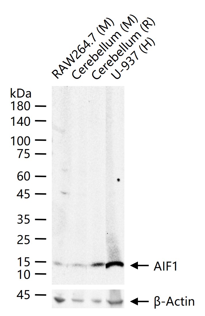
AIF1/Iba1 Rabbit pAb
AIF1/Iba1 Rabbit pAb
- SPECIFICATION
- CITATIONS
- PROTOCOLS
- BACKGROUND

Application
| WB, IHC-P, IHC-F, E |
|---|---|
| Primary Accession | O70200 |
| Reactivity | Rat, Human, Mouse |
| Host | Rabbit |
| Clonality | Polyclonal |
| Calculated MW | 16 KDa |
| Physical State | Liquid |
| Immunogen | KLH conjugated synthetic peptide derived from human Iba1 |
| Epitope Specificity | 51-147/147 |
| Isotype | IgG |
| Purity | affinity purified by Protein A |
| Buffer | 0.01M TBS (pH7.4) with 1% BSA, 0.02% Proclin300 and 50% Glycerol. |
| SUBCELLULAR LOCATION | Cytoplasm, cytoskeleton. Cell projection, ruffle membrane; Peripheral membrane protein; Cytoplasmic side. Note=Associated with the actin cytoskeleton at membrane ruffles and at sites of phagocytosis. |
| SIMILARITY | Contains 2 EF-hand domains. |
| SUBUNIT | Homodimer (Potential). Monomer. Interacts with LCP1. |
| Important Note | This product as supplied is intended for research use only, not for use in human, therapeutic or diagnostic applications. |
| Background Descriptions | Allograft Inflammatory Factor-1 (AIF1)or ionized calcium-binding adaptor molecule 1 (Iba1) is expressed selectively in microglia/macrophages and is a Ca2+-binding peptide produced by activated monocytes and microglial cells. It has been suggested that AIF1 expression is associated with chronic inflammatory processes. AIF1 is expressed by activated monocytes and might participate in a variety of pathogenic processes in the mammalian brain and in chronic transplant rejection. It has been shown to be expressed early and persistently in chronically rejecting cardiac allografts but not in cardiac syngrafts and host hearts. |
| Gene ID | 11629 |
|---|---|
| Other Names | Allograft inflammatory factor 1, AIF-1, Ionized calcium-binding adapter molecule 1, Aif1, Iba1 |
| Target/Specificity | Detected in T-lymphocytes and peripheral blood mononuclear cells. |
| Dilution | WB=1:500-2000,IHC-P=1:100-500,IHC-F=1:100-500,Flow-Cyt=0.2ug/test,ELISA=1:5000-10000 |
| Format | 0.01M TBS(pH7.4), 0.09% (W/V) sodium azide and 50% Glyce |
| Storage | Store at -20 ℃ for one year. Avoid repeated freeze/thaw cycles. When reconstituted in sterile pH 7.4 0.01M PBS or diluent of antibody the antibody is stable for at least two weeks at 2-4 ℃. |
| Name | Aif1 |
|---|---|
| Synonyms | Iba1 |
| Function | Actin-binding protein that enhances membrane ruffling and RAC activation. Enhances the actin-bundling activity of LCP1. Binds calcium. Plays a role in RAC signaling and in phagocytosis. May play a role in macrophage activation and function. Promotes the proliferation of vascular smooth muscle cells and of T-lymphocytes. Enhances lymphocyte migration. Plays a role in vascular inflammation. |
| Cellular Location | Cytoplasm, cytoskeleton. Cell projection, ruffle membrane; Peripheral membrane protein; Cytoplasmic side. Cell projection, phagocytic cup Note=Associated with the actin cytoskeleton at membrane ruffles and at sites of phagocytosis. |
| Tissue Location | Abundantly expressed in the testis, moderately in the spleen and lymph nodes and at low levels in the liver and thymus Detected in macrophages. |

Thousands of laboratories across the world have published research that depended on the performance of antibodies from Abcepta to advance their research. Check out links to articles that cite our products in major peer-reviewed journals, organized by research category.
info@abcepta.com, and receive a free "I Love Antibodies" mug.

Provided below are standard protocols that you may find useful for product applications.
Background
This product as supplied is intended for research use only, not for use in human, therapeutic or diagnostic applications.
If you have used an Abcepta product and would like to share how it has performed, please click on the "Submit Review" button and provide the requested information. Our staff will examine and post your review and contact you if needed.
If you have any additional inquiries please email technical services at tech@abcepta.com.
Ordering Information
Other Products
Shipping Information
