


 Foundational characteristics of cancer include proliferation, angiogenesis, migration, evasion of apoptosis, and cellular immortality. Find key markers for these cellular processes and antibodies to detect them.
 The SUMOplot™ Analysis Program predicts and scores sumoylation sites in your protein. SUMOylation is a post-translational modification involved in various cellular processes, such as nuclear-cytosolic transport, transcriptional regulation, apoptosis, protein stability, response to stress, and progression through the cell cycle.
 The Autophagy Receptor Motif Plotter predicts and scores autophagy receptor binding sites in your protein. Identifying proteins connected to this pathway is critical to understanding the role of autophagy in physiological as well as pathological processes such as development, differentiation, neurodegenerative diseases, stress, infection, and cancer.
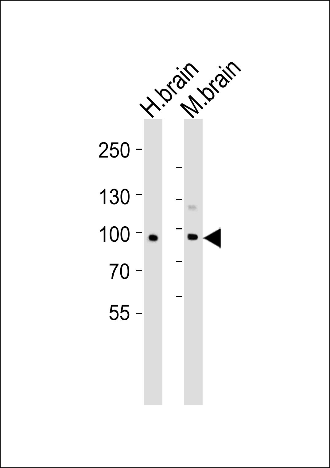
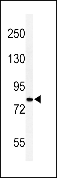
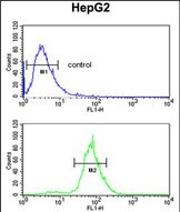
CDH20 Antibody (N-term)
Affinity Purified Rabbit Polyclonal Antibody (Pab)
- SPECIFICATION
- CITATIONS
- PROTOCOLS
- BACKGROUND

Application
| WB, FC, E |
|---|---|
| Primary Accession | Q9HBT6 |
| Other Accession | Q5DWV1, Q9Z0M3 |
| Reactivity | Human |
| Predicted | Mouse, Rat |
| Host | Rabbit |
| Clonality | Polyclonal |
| Isotype | Rabbit IgG |
| Calculated MW | 88993 Da |
| Antigen Region | 111-140 aa |
| Gene ID | 28316 |
|---|---|
| Other Names | Cadherin-20, CDH20, CDH7L3 |
| Target/Specificity | This CDH20 antibody is generated from rabbits immunized with a KLH conjugated synthetic peptide between 111-140 amino acids from the N-terminal region of human CDH20. |
| Dilution | WB~~1:1000 FC~~1:10~50 E~~Use at an assay dependent concentration. |
| Format | Purified polyclonal antibody supplied in PBS with 0.09% (W/V) sodium azide. This antibody is purified through a protein A column, followed by peptide affinity purification. |
| Storage | Maintain refrigerated at 2-8°C for up to 2 weeks. For long term storage store at -20°C in small aliquots to prevent freeze-thaw cycles. |
| Precautions | CDH20 Antibody (N-term) is for research use only and not for use in diagnostic or therapeutic procedures. |
| Name | CDH20 |
|---|---|
| Synonyms | CDH7L3 |
| Function | Cadherins are calcium-dependent cell adhesion proteins. They preferentially interact with themselves in a homophilic manner in connecting cells; cadherins may thus contribute to the sorting of heterogeneous cell types. |
| Cellular Location | Cell membrane; Single-pass type I membrane protein |
| Tissue Location | Expressed in placenta, adult brain, and fetal brain |

Thousands of laboratories across the world have published research that depended on the performance of antibodies from Abcepta to advance their research. Check out links to articles that cite our products in major peer-reviewed journals, organized by research category.
info@abcepta.com, and receive a free "I Love Antibodies" mug.

Provided below are standard protocols that you may find useful for product applications.
Background
This gene is a type II classical cadherin from the cadherin superfamily and one of three cadherin 7-like genes located in a cluster on chromosome 18. The encoded membrane protein is a calcium dependent cell-cell adhesion glycoprotein comprised of five extracellular cadherin repeats, a transmembrane region and a highly conserved cytoplasmic tail. Type II (atypical) cadherins are defined based on their lack of a HAV cell adhesion recognition sequence specific to type I cadherins. Since disturbance of intracellular adhesion is a prerequisite for invasion and metastasis of tumor cells, cadherins are considered prime candidates for tumor suppressor genes.
References
Kools, P., et al. Genomics 68(3):283-295(2000)
If you have used an Abcepta product and would like to share how it has performed, please click on the "Submit Review" button and provide the requested information. Our staff will examine and post your review and contact you if needed.
If you have any additional inquiries please email technical services at tech@abcepta.com.
Ordering Information
Other Products
Shipping Information
