


 Foundational characteristics of cancer include proliferation, angiogenesis, migration, evasion of apoptosis, and cellular immortality. Find key markers for these cellular processes and antibodies to detect them.
 The SUMOplot™ Analysis Program predicts and scores sumoylation sites in your protein. SUMOylation is a post-translational modification involved in various cellular processes, such as nuclear-cytosolic transport, transcriptional regulation, apoptosis, protein stability, response to stress, and progression through the cell cycle.
 The Autophagy Receptor Motif Plotter predicts and scores autophagy receptor binding sites in your protein. Identifying proteins connected to this pathway is critical to understanding the role of autophagy in physiological as well as pathological processes such as development, differentiation, neurodegenerative diseases, stress, infection, and cancer.
Bcl-B Antibody
- SPECIFICATION
- CITATIONS
- PROTOCOLS
- BACKGROUND

Application
| WB, ICC, E |
|---|---|
| Primary Accession | Q9HD36 |
| Other Accession | NP_065129, 10017 |
| Reactivity | Human |
| Host | Rabbit |
| Clonality | Polyclonal |
| Isotype | IgG |
| Calculated MW | 23204 Da |
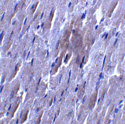
| Application Notes | Bcl-B antibody can be used for the detection of Bcl-B by Western blot at 1 - 2 μg/mL. Despite its predicted molecular weight, Bcl-B is often at higher molecular weights, presumably due to post-translational modifications. Antibody can also be used for immunocytochemistry starting at 10 μg/mL. |
| Gene ID | 10017 |
|---|---|
| Other Names | Bcl-B Antibody: Boo, Diva, BCL-B, BCLB, Bcl-2-like protein 10, Anti-apoptotic protein NrH, Bcl2-L-10, BCL2-like 10 (apoptosis facilitator) |
| Target/Specificity | Bcl-B antibody was raised against a 16 amino acid synthetic peptide from near the amino terminus of human Bcl-B. The immunogen is located within amino acids 90 - 140 of Bcl-B. |
| Reconstitution & Storage | Bcl-B antibody can be stored at 4℃ for three months and -20℃, stable for up to one year. As with all antibodies care should be taken to avoid repeated freeze thaw cycles. Antibodies should not be exposed to prolonged high temperatures. |
| Precautions | Bcl-B Antibody is for research use only and not for use in diagnostic or therapeutic procedures. |
| Name | BCL2L10 {ECO:0000303|PubMed:17532299} |
|---|---|
| Function | Promotes cell survival by suppressing apoptosis induced by BAX but not BAK (PubMed:11278245, PubMed:11689480). Increases binding of AHCYL1/IRBIT to ITPR1 (PubMed:27995898). Reduces ITPR1-mediated calcium release from the endoplasmic reticulum cooperatively with AHCYL1/IRBIT under normal cellular conditions (PubMed:27995898). Under apoptotic stress conditions, dissociates from ITPR1 and is displaced from mitochondria-associated endoplasmic reticulum membranes, leading to increased Ca(2+) transfer to mitochondria which promotes apoptosis (PubMed:27995898). Required for the correct formation of the microtubule organizing center during oocyte cell division, potentially via regulation of protein abundance and localization of other microtubule organizing center components such as AURKA and TPX2 (By similarity). |
| Cellular Location | Mitochondrion. Nucleus membrane. Endoplasmic reticulum. Cytoplasm, cytoskeleton, spindle {ECO:0000250|UniProtKB:Q9Z0F3}. Note=Localizes to mitochondria-associated endoplasmic reticulum membranes (MAMs) (PubMed:27995898). Localization to MAMs is greatly reduced under apoptotic stress conditions (PubMed:27995898) |
| Tissue Location | Widely expressed in adult tissues. Preferentially expressed in lung, liver and kidney. |

Thousands of laboratories across the world have published research that depended on the performance of antibodies from Abcepta to advance their research. Check out links to articles that cite our products in major peer-reviewed journals, organized by research category.
info@abcepta.com, and receive a free "I Love Antibodies" mug.

Provided below are standard protocols that you may find useful for product applications.
Background
Bcl-B Antibody: Members in the Bcl-2 family are critical regulators of apoptosis by either inhibiting or promoting cell death. Bcl-B is a recently discovered anti-apoptotic member of the Bcl-2. Unlike the mouse homolog (also known as Diva/Boo) which is predominantly expressed in ovary and testis, the human Bcl-B protein is widely expressed. Also, the human Bcl-B functions by binding to and suppressing the apoptotic activity of Bax, whereas the mouse homolog binds Bak and also interacts with the apoptosis protein Apaf-1.
References
Cory S, Huang DCS, and Adams JM. The Bcl-2 family: roles in cell survival and oncogenesis. Oncogene 2003; 22:8590-607
Heiser D, Labi V, Erlacher M, et al. The Bcl-2 protein family and its role in the development of neoplastic disease. Exp. Geron. 2004; 39:1125-35.
Ke N, Godzik A, and Reed JC. Bcl-B: A novel Bcl-2 family member that differentially binds and regulates Bax and Bak. J. Biol. Chem. 2001; 276:12481-4.
Song Q, Kuang Y, Dixit VM, et al. Boo, a negative regulator of cell death, interacts with Apaf-1. EMBO J. 1999; 18:167-78.
If you have used an Abcepta product and would like to share how it has performed, please click on the "Submit Review" button and provide the requested information. Our staff will examine and post your review and contact you if needed.
If you have any additional inquiries please email technical services at tech@abcepta.com.
Ordering Information
Shipping Information
