


 Foundational characteristics of cancer include proliferation, angiogenesis, migration, evasion of apoptosis, and cellular immortality. Find key markers for these cellular processes and antibodies to detect them.
 The SUMOplot™ Analysis Program predicts and scores sumoylation sites in your protein. SUMOylation is a post-translational modification involved in various cellular processes, such as nuclear-cytosolic transport, transcriptional regulation, apoptosis, protein stability, response to stress, and progression through the cell cycle.
 The Autophagy Receptor Motif Plotter predicts and scores autophagy receptor binding sites in your protein. Identifying proteins connected to this pathway is critical to understanding the role of autophagy in physiological as well as pathological processes such as development, differentiation, neurodegenerative diseases, stress, infection, and cancer.
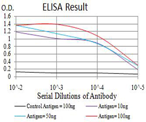
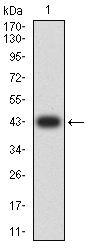
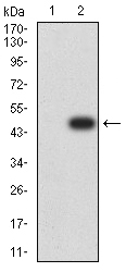
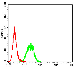
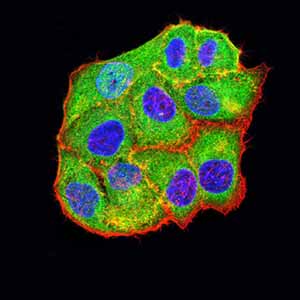
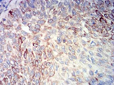
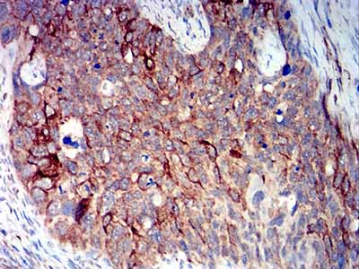
BCL2L10
Purified Mouse Monoclonal Antibody
- SPECIFICATION
- CITATIONS
- PROTOCOLS
- BACKGROUND

Application
| WB, IHC, ICC, E |
|---|---|
| Primary Accession | Q9HD36 |
| Reactivity | Human |
| Host | Mouse |
| Clonality | Monoclonal |
| Clone Names | 8A2F9 |
| Isotype | Mouse IgG1 |
| Calculated MW | 22kDa |
| Immunogen | Purified recombinant fragment of human BCL2L10 (AA: 31-186) expressed in E. Coli. |
| Formulation | Purified antibody in PBS with 0.05% sodium azide |
| Gene ID | 10017 |
|---|---|
| Other Names | Boo; Diva; BCL-B; bcl2-L-10 |
| Dilution | WB~~ 1/500 - 1/2000 IHC~~ 1/200 - 1/1000 ICC~~ 1/200 - 1/1000 E~~ 1/10000 |
| Storage | Maintain refrigerated at 2-8°C for up to 6 months. For long term storage store at -20°C in small aliquots to prevent freeze-thaw cycles. |
| Precautions | BCL2L10 is for research use only and not for use in diagnostic or therapeutic procedures. |
| Name | BCL2L10 {ECO:0000303|PubMed:17532299} |
|---|---|
| Function | Promotes cell survival by suppressing apoptosis induced by BAX but not BAK (PubMed:11278245, PubMed:11689480). Increases binding of AHCYL1/IRBIT to ITPR1 (PubMed:27995898). Reduces ITPR1-mediated calcium release from the endoplasmic reticulum cooperatively with AHCYL1/IRBIT under normal cellular conditions (PubMed:27995898). Under apoptotic stress conditions, dissociates from ITPR1 and is displaced from mitochondria-associated endoplasmic reticulum membranes, leading to increased Ca(2+) transfer to mitochondria which promotes apoptosis (PubMed:27995898). Required for the correct formation of the microtubule organizing center during oocyte cell division, potentially via regulation of protein abundance and localization of other microtubule organizing center components such as AURKA and TPX2 (By similarity). |
| Cellular Location | Mitochondrion. Nucleus membrane. Endoplasmic reticulum. Cytoplasm, cytoskeleton, spindle {ECO:0000250|UniProtKB:Q9Z0F3}. Note=Localizes to mitochondria-associated endoplasmic reticulum membranes (MAMs) (PubMed:27995898). Localization to MAMs is greatly reduced under apoptotic stress conditions (PubMed:27995898) |
| Tissue Location | Widely expressed in adult tissues. Preferentially expressed in lung, liver and kidney. |

Thousands of laboratories across the world have published research that depended on the performance of antibodies from Abcepta to advance their research. Check out links to articles that cite our products in major peer-reviewed journals, organized by research category.
info@abcepta.com, and receive a free "I Love Antibodies" mug.

Provided below are standard protocols that you may find useful for product applications.
References
1.Hum Reprod. 2013 Mar;28(3):729-39. 2.Oncotarget. 2012 Apr;3(4):490-501.
If you have used an Abcepta product and would like to share how it has performed, please click on the "Submit Review" button and provide the requested information. Our staff will examine and post your review and contact you if needed.
If you have any additional inquiries please email technical services at tech@abcepta.com.
Ordering Information
Shipping Information
