


 Foundational characteristics of cancer include proliferation, angiogenesis, migration, evasion of apoptosis, and cellular immortality. Find key markers for these cellular processes and antibodies to detect them.
 The SUMOplot™ Analysis Program predicts and scores sumoylation sites in your protein. SUMOylation is a post-translational modification involved in various cellular processes, such as nuclear-cytosolic transport, transcriptional regulation, apoptosis, protein stability, response to stress, and progression through the cell cycle.
 The Autophagy Receptor Motif Plotter predicts and scores autophagy receptor binding sites in your protein. Identifying proteins connected to this pathway is critical to understanding the role of autophagy in physiological as well as pathological processes such as development, differentiation, neurodegenerative diseases, stress, infection, and cancer.
SYNGR1 Antibody
- SPECIFICATION
- CITATIONS
- PROTOCOLS
- BACKGROUND

Application
| WB, IHC-P, IF, E |
|---|---|
| Primary Accession | O43759 |
| Other Accession | CAA05322, 2959866 |
| Reactivity | Human, Mouse, Rat |
| Host | Rabbit |
| Clonality | Polyclonal |
| Isotype | IgG |
| Calculated MW | 25456 Da |
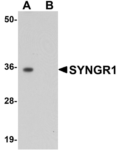
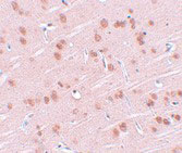
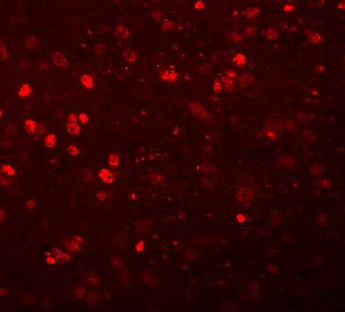
| Application Notes | SYNGR1 antibody can be used for detection of SYNGR1 by Western blot at 1 µg/mL. Antibody can also be used for immunohistochemistry starting at 2.5 µg/mL. For immunofluorescence start at 20 µg/mL. |
| Gene ID | 9145 |
|---|---|
| Target/Specificity | SYNGR1; |
| Reconstitution & Storage | Antibody can be stored at 4°C up to one year. Antibodies should not be exposed to prolonged high temperatures. |
| Precautions | SYNGR1 Antibody is for research use only and not for use in diagnostic or therapeutic procedures. |
| Name | SYNGR1 (HGNC:11498) |
|---|---|
| Function | May play a role in regulated exocytosis. Modulates the localization of synaptophysin/SYP into synaptic-like microvesicles and may therefore play a role in synaptic-like microvesicle formation and/or maturation (By similarity). Involved in the regulation of short- term and long-term synaptic plasticity (By similarity). |
| Cellular Location | Cytoplasmic vesicle, secretory vesicle, synaptic vesicle membrane {ECO:0000250|UniProtKB:Q62876}; Multi-pass membrane protein {ECO:0000250|UniProtKB:Q62876}. Melanosome. Note=Identified by mass spectrometry in melanosome fractions from stage I to stage IV |

Thousands of laboratories across the world have published research that depended on the performance of antibodies from Abcepta to advance their research. Check out links to articles that cite our products in major peer-reviewed journals, organized by research category.
info@abcepta.com, and receive a free "I Love Antibodies" mug.

Provided below are standard protocols that you may find useful for product applications.
Background
SYNGR1 Antibody: Synaptogyrins comprise a family of tyrosine-phosphorylated membrane proteins with two neuronal (SYNGR1 and SYNGR3) and one ubiquitous (SYNGR2) members. SYNGR1 and -3 are synaptic vesicle proteins, residing in some cases on the same synaptic vesicle, and are thought to be involved in the regulation of neurotransmitter release. SYNGR2, by contrast, is absent from synaptic vesicles. The role and localization of a fourth synaptogyrin, SYNGR4, is unclear. The gene for SYNGR1is located at chromosome 22q13, a region linked to schizophrenia; however, there is mixed evidence suggesting that mutations in SYNGR1 might be associated with schizophrenia.
References
Kedra D, Pan HQ, Seroussi E, et al. Characterization of the human synaptogyrin gene family. Hum. Genet.1998; 103:131-41.
Egaña LA, Cuevas RA, Baust TB, et al. Physical and functional interaction between the dopamine transporter and the synaptic vesicle protein synaptogyrin-3. J. Neurosci.2009; 29:4592-604.
Cheng MC and Chen CH. Identification of rare mutations of synaptogyrin 1 gene in patients with schizophrenia. J. Psychiatr. Res.2007; 41:1027-31.
Wang Y, Yu L, Zhao T, et al. No association between bipolar disorder and syngr1 or synapsin II polymorphisms in the Han Chinese population. Psychiatry Res.2009; 169:167-8.
If you have used an Abcepta product and would like to share how it has performed, please click on the "Submit Review" button and provide the requested information. Our staff will examine and post your review and contact you if needed.
If you have any additional inquiries please email technical services at tech@abcepta.com.
Ordering Information
Other Products
Shipping Information
