


 Foundational characteristics of cancer include proliferation, angiogenesis, migration, evasion of apoptosis, and cellular immortality. Find key markers for these cellular processes and antibodies to detect them.
 The SUMOplot™ Analysis Program predicts and scores sumoylation sites in your protein. SUMOylation is a post-translational modification involved in various cellular processes, such as nuclear-cytosolic transport, transcriptional regulation, apoptosis, protein stability, response to stress, and progression through the cell cycle.
 The Autophagy Receptor Motif Plotter predicts and scores autophagy receptor binding sites in your protein. Identifying proteins connected to this pathway is critical to understanding the role of autophagy in physiological as well as pathological processes such as development, differentiation, neurodegenerative diseases, stress, infection, and cancer.
TRIP6 Antibody


- SPECIFICATION
- CITATIONS
- PROTOCOLS
- BACKGROUND

Application
| WB, IF, ICC, E |
|---|---|
| Primary Accession | Q15654 |
| Other Accession | NP_003293, 91208423 |
| Reactivity | Human, Mouse |
| Host | Rabbit |
| Clonality | Polyclonal |
| Isotype | IgG |
| Calculated MW | 50288 Da |
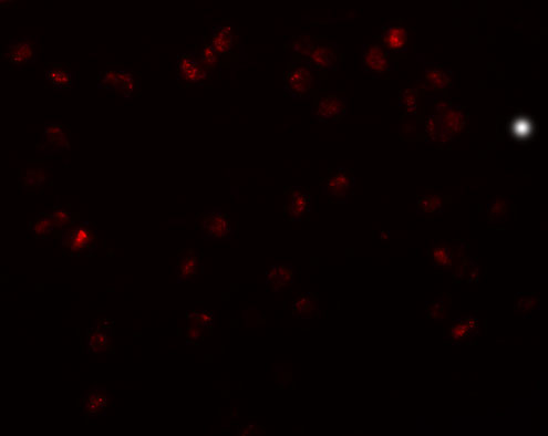
| Application Notes | TRIP6 antibody can be used for detection of TRIP6 by Western blot at 1 µg/mL. Antibody can also be used for immunocytochemistry starting at µg/mL. For immunofluorescence start at 20 µg/mL. |
| Gene ID | 7205 |
|---|---|
| Target/Specificity | TRIP6; |
| Reconstitution & Storage | TRIP6 antibody can be stored at 4℃ for three months and -20℃, stable for up to one year. As with all antibodies care should be taken to avoid repeated freeze thaw cycles. Antibodies should not be exposed to prolonged high temperatures. |
| Precautions | TRIP6 Antibody is for research use only and not for use in diagnostic or therapeutic procedures. |
| Name | TRIP6 |
|---|---|
| Synonyms | OIP1 |
| Function | Relays signals from the cell surface to the nucleus to weaken adherens junction and promote actin cytoskeleton reorganization and cell invasiveness. Involved in lysophosphatidic acid-induced cell adhesion and migration. Acts as a transcriptional coactivator for NF- kappa-B and JUN, and mediates the transrepression of these transcription factors induced by glucocorticoid receptor. |
| Cellular Location | Cytoplasm, cytoskeleton. Cell junction, focal adhesion. Nucleus. Cytoplasm Note=Shuttles between nucleus and cytoplasm (PubMed:16624523) Colocalizes with actin (PubMed:10826496). |
| Tissue Location | Abundantly expressed in kidney, liver and lung. Lower levels in heart, placenta and pancreas. Expressed in colonic epithelial cells. Up-regulated in colonic tumors |

Thousands of laboratories across the world have published research that depended on the performance of antibodies from Abcepta to advance their research. Check out links to articles that cite our products in major peer-reviewed journals, organized by research category.
info@abcepta.com, and receive a free "I Love Antibodies" mug.

Provided below are standard protocols that you may find useful for product applications.
Background
TRIP6 Antibody: TRIP6 (Thyroid receptor interacting protein 6) is a zyxin-related focal adhesion molecule involved in cell motility and antiapoptotic responses. TRIP6 is predominantly expressed in kidney, liver and lung. It has three LIM domains, a PDZ-binding motif, a Crk SH2-binding motif and/or other protein interacting domains. TRIP6 regulates NF-κB activation and interacts with RICK and TRAF2, proteins that are involved in TNF signaling. TRIP6 antagonizes Fas-induced apoptosis and promotes Fas-mediated cell migration in apoptosis-resistant glioma cells via Src-dependent phosphorylation of TRIP6 at Tyr-55.
References
Murthy KK, Clark K, Fortin Y, et al. ZRP-1, a zyxin-related protein, interacts with the second PDZ domain of the cytosolic protein tyrosine phosphatase hPTP1E. J. Biol. Chem.1999; 274:20679-87.
Kassel O, Schneider S, Heilbock C, et al. A nuclear isoform of the focal adhesion LIM-domain protein Trip6 integrates activating and repressing signals at AP-1- and NF-kappaB regulated promoters. Genes Dev.2004; 18:2518-2528.
Lai YJ, Chen CS, Lin WJ, et al. c-Src-mediated phosphorylation of TRIP6 regulates its function in lysophosphatidic acid-induced cell migration. Mol. Cell. Biol.2005; 25:5859-5868.
Li L, Bin LH, Li F, et al. TRIP6 is a RIP2-associated common signaling component of multiple NF-kB activation pathways. J. Cell. Sci.2005; 118:555-563.
If you have used an Abcepta product and would like to share how it has performed, please click on the "Submit Review" button and provide the requested information. Our staff will examine and post your review and contact you if needed.
If you have any additional inquiries please email technical services at tech@abcepta.com.
Ordering Information
Other Products
Shipping Information
