


 Foundational characteristics of cancer include proliferation, angiogenesis, migration, evasion of apoptosis, and cellular immortality. Find key markers for these cellular processes and antibodies to detect them.
 The SUMOplot™ Analysis Program predicts and scores sumoylation sites in your protein. SUMOylation is a post-translational modification involved in various cellular processes, such as nuclear-cytosolic transport, transcriptional regulation, apoptosis, protein stability, response to stress, and progression through the cell cycle.
 The Autophagy Receptor Motif Plotter predicts and scores autophagy receptor binding sites in your protein. Identifying proteins connected to this pathway is critical to understanding the role of autophagy in physiological as well as pathological processes such as development, differentiation, neurodegenerative diseases, stress, infection, and cancer.
EIG121 Antibody
- SPECIFICATION
- CITATIONS
- PROTOCOLS
- BACKGROUND

Application
| WB, IF, ICC, E |
|---|---|
| Primary Accession | Q6UXG2 |
| Other Accession | NP_065826, 38569482 |
| Reactivity | Human |
| Host | Rabbit |
| Clonality | Polyclonal |
| Isotype | IgG |
| Calculated MW | 111382 Da |
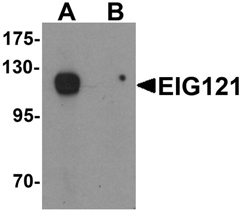
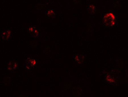
| Application Notes | EIG121 antibody can be used for detection of EIG121 by Western blot at 1 µg/mL. Antibody can also be used for immunocytochemistry starting at 5 µg/mL. For immunofluorescence start at 20 µg/mL. |
| Gene ID | 1957 |
|---|---|
| Target/Specificity | EGI; At least four isoforms of EIG121 known to exist. EIG121 antibody is predicted to not cross-react with other UPF0577 protein family members. |
| Reconstitution & Storage | EIG121 antibody can be stored at 4℃ for three months and -20℃, stable for up to one year. As with all antibodies care should be taken to avoid repeated freeze thaw cycles. Antibodies should not be exposed to prolonged high temperatures. |
| Precautions | EIG121 Antibody is for research use only and not for use in diagnostic or therapeutic procedures. |
| Name | ELAPOR1 (HGNC:29618) |
|---|---|
| Function | May protect cells from cell death by inducing cytosolic vacuolization and up-regulating the autophagy pathway (PubMed:21072319). May play a role in apoptosis and cell proliferation through its interaction with HSPA5 (PubMed:26045166). |
| Cellular Location | Cell membrane; Single-pass type I membrane protein. Late endosome membrane; Single-pass type I membrane protein. Golgi apparatus, trans-Golgi network membrane; Single-pass type I membrane protein. Lysosome membrane; Single-pass membrane protein. Endoplasmic reticulum membrane; Single-pass type I membrane protein |
| Tissue Location | Expressed in normal endometrium but overexpressed in endometroid tumors. |

Thousands of laboratories across the world have published research that depended on the performance of antibodies from Abcepta to advance their research. Check out links to articles that cite our products in major peer-reviewed journals, organized by research category.
info@abcepta.com, and receive a free "I Love Antibodies" mug.

Provided below are standard protocols that you may find useful for product applications.
Background
EIG121 Antibody: EIG121 (Estrogen-induced gene 121 protein) is thought to play a role as a marker of hyperestrogenic state and estrogen-related type I endometrial carcinoma. It belongs to the UPF0577 family. It is a 1013 amino acid single-pass transmembrane protein that, though expressed in normal endometrium, is overexpressed in endometriod tumors. As an evolutionarily conserved gene, EIG121 is also expressed during early xenopus development, showing maximum expression at the gastrula stage.
References
Deng L, Broaddus RR, McCampbell A, et al. Identification of a novel estrogen-regulated gene, EIG121, induced by hormone replacement therapy and differentially expressed in type I and type II endometrial cancer. Clin. Cancer Res. 2005; 11:8258-64.
Deng L, Feng J, Broaddus RR. The novel estrogen-induced gene EIG121 regulates autophagy and promotes cell survival under stress. Cell Death Dis. 2010; 1:e32.
Araki T, Kusakabe M and Nishida E. Expression of estrogen induced gene 121-like (EIG121L) during early Xenopus development. Gene Expr. Patterns 2007; 7:666-71.
If you have used an Abcepta product and would like to share how it has performed, please click on the "Submit Review" button and provide the requested information. Our staff will examine and post your review and contact you if needed.
If you have any additional inquiries please email technical services at tech@abcepta.com.
Ordering Information
Other Products
Shipping Information
