


 Foundational characteristics of cancer include proliferation, angiogenesis, migration, evasion of apoptosis, and cellular immortality. Find key markers for these cellular processes and antibodies to detect them.
 The SUMOplot™ Analysis Program predicts and scores sumoylation sites in your protein. SUMOylation is a post-translational modification involved in various cellular processes, such as nuclear-cytosolic transport, transcriptional regulation, apoptosis, protein stability, response to stress, and progression through the cell cycle.
 The Autophagy Receptor Motif Plotter predicts and scores autophagy receptor binding sites in your protein. Identifying proteins connected to this pathway is critical to understanding the role of autophagy in physiological as well as pathological processes such as development, differentiation, neurodegenerative diseases, stress, infection, and cancer.
JMJD3 Antibody
- SPECIFICATION
- CITATIONS
- PROTOCOLS
- BACKGROUND

Application
| WB, IF, ICC, E |
|---|---|
| Primary Accession | O15054 |
| Other Accession | EAW90126, 122937251 |
| Reactivity | Human, Mouse, Rat |
| Host | Rabbit |
| Clonality | Polyclonal |
| Isotype | IgG |
| Calculated MW | 176632 Da |
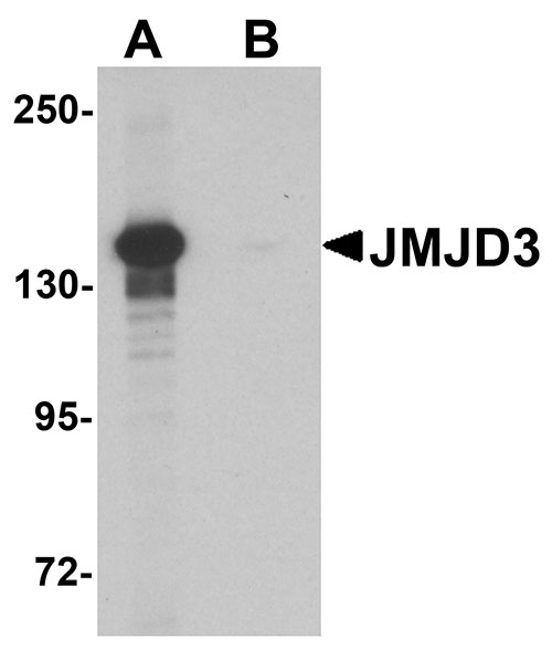
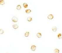
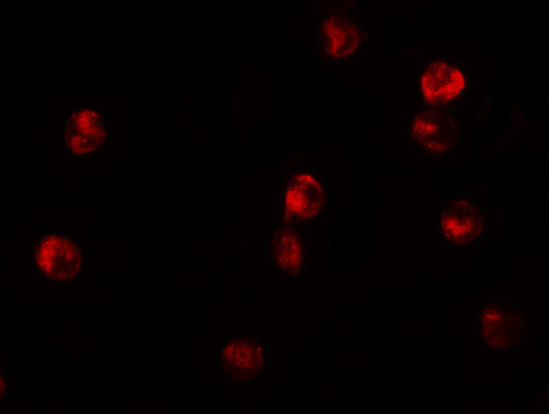
| Application Notes | JMJD3 antibody can be used for detection of EPAC1 by Western blot at 0.5 µg/mL. Antibody can also be used for immunocytochemistry starting at 2.5 µg/mL. For immunofluorescence start at 5 µg/mL. |
| Gene ID | 23135 |
|---|---|
| Target/Specificity | KDM6B; This antibody is specific for JMJD3 and will not recognize other JMJD proteins. |
| Reconstitution & Storage | JMJD3 antibody can be stored at 4℃ for three months and -20℃, stable for up to one year. As with all antibodies care should be taken to avoid repeated freeze thaw cycles. Antibodies should not be exposed to prolonged high temperatures. |
| Precautions | JMJD3 Antibody is for research use only and not for use in diagnostic or therapeutic procedures. |
| Name | KDM6B |
|---|---|
| Synonyms | JMJD3, KIAA0346 |
| Function | Histone demethylase that specifically demethylates 'Lys-27' of histone H3, thereby playing a central role in histone code (PubMed:17713478, PubMed:17825402, PubMed:17851529, PubMed:18003914). Demethylates trimethylated and dimethylated H3 'Lys-27' (PubMed:17713478, PubMed:17825402, PubMed:17851529, PubMed:18003914). Plays a central role in regulation of posterior development, by regulating HOX gene expression (PubMed:17851529). Involved in inflammatory response by participating in macrophage differentiation in case of inflammation by regulating gene expression and macrophage differentiation (PubMed:17825402). Plays a demethylase-independent role in chromatin remodeling to regulate T-box family member-dependent gene expression by acting as a link between T-box factors and the SMARCA4- containing SWI/SNF remodeling complex (By similarity). |
| Cellular Location | Nucleus. |

Thousands of laboratories across the world have published research that depended on the performance of antibodies from Abcepta to advance their research. Check out links to articles that cite our products in major peer-reviewed journals, organized by research category.
info@abcepta.com, and receive a free "I Love Antibodies" mug.

Provided below are standard protocols that you may find useful for product applications.
Background
JMJD3 Antibody: The Jumonji domain-containing protein 3 (JMJD3) functions as a trimethylation-specific demethylase, converting the trimethylated histone H3 Lys27 residue to the dimethylated form, and is thought to also function as a transcriptional repressor. JMJD3 plays a central role in regulation of posterior development, by regulating HOX gene expression. It is involved in inflammatory response by participating in macrophage differentiation in case of inflammation by regulating gene expression and macrophage differentiation. JMJD3 can also interact with and demethylate p53, resulting in its stabilization and localization to the nucleus in mouse embryo fibroblasts during neural stem cell differentiation.
References
Agger K, Cloos PA, Christensen J, et al. UTX and JMJD3 are histone H3K27 demethylases involved in HOX gene regulation and development. Nature 2007; 449:731-4.
De Santa F, Totaro MG, Prosperini E, et al. The histone H3 lysine-27 demethylase Jmjd3 links inflammation to inhibition of polycomb-mediated silencing. Cell 2007; 130:1083-94
Sola S, Xavier JM, Santos DM, et al. p53 interaction with JMJD3 results in its nuclear distribution during mouse neural stem cell differentiation. PLoS One 2011; 6:e18421.
If you have used an Abcepta product and would like to share how it has performed, please click on the "Submit Review" button and provide the requested information. Our staff will examine and post your review and contact you if needed.
If you have any additional inquiries please email technical services at tech@abcepta.com.
Ordering Information
Other Products
Shipping Information
