


 Foundational characteristics of cancer include proliferation, angiogenesis, migration, evasion of apoptosis, and cellular immortality. Find key markers for these cellular processes and antibodies to detect them.
 The SUMOplot™ Analysis Program predicts and scores sumoylation sites in your protein. SUMOylation is a post-translational modification involved in various cellular processes, such as nuclear-cytosolic transport, transcriptional regulation, apoptosis, protein stability, response to stress, and progression through the cell cycle.
 The Autophagy Receptor Motif Plotter predicts and scores autophagy receptor binding sites in your protein. Identifying proteins connected to this pathway is critical to understanding the role of autophagy in physiological as well as pathological processes such as development, differentiation, neurodegenerative diseases, stress, infection, and cancer.
TTYH1 Antibody
- SPECIFICATION
- CITATIONS
- PROTOCOLS
- BACKGROUND

Application
| WB, ICC, E |
|---|---|
| Primary Accession | Q9H313 |
| Other Accession | NP_001005367, 53831989 |
| Reactivity | Human |
| Host | Rabbit |
| Clonality | Polyclonal |
| Isotype | IgG |
| Calculated MW | 49051 Da |
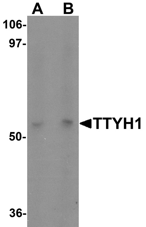
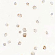
| Application Notes | TTYH1 antibody can be used for detection of TTYH1 by Western blot at 1 - 2 µg/mL. Antibody can also be used for immunocytochemistry starting at 5 µg/mL. |
| Gene ID | 57348 |
|---|---|
| Target/Specificity | TTYH1; TTYH1 antibody is human specific. Multiple isoforms of TTYH1 are known to exist. |
| Reconstitution & Storage | TTYH1 antibody can be stored at 4℃ for three months and -20℃, stable for up to one year. As with all antibodies care should be taken to avoid repeated freeze thaw cycles. Antibodies should not be exposed to prolonged high temperatures. |
| Precautions | TTYH1 Antibody is for research use only and not for use in diagnostic or therapeutic procedures. |
| Name | TTYH1 |
|---|---|
| Function | Calcium-independent, swelling-dependent volume-regulated anion channel (VRAC-swell) which plays a pivotal role in the process of regulatory volume decrease (RVD) in the brain through the efflux of anions like chloride and organic osmolytes like glutamate. |
| Cellular Location | Cell membrane; Multi-pass membrane protein |
| Tissue Location | Expressed in brain, eye, ovary and testis, and at lower levels in muscle, placenta, liver and lung |

Thousands of laboratories across the world have published research that depended on the performance of antibodies from Abcepta to advance their research. Check out links to articles that cite our products in major peer-reviewed journals, organized by research category.
info@abcepta.com, and receive a free "I Love Antibodies" mug.

Provided below are standard protocols that you may find useful for product applications.
Background
TTYH1 Antibody: TTYH1 is a member of the tweety family of proteins, a family of chloride anion channels containing five transmembrane regions. TTYH1 is a Ca2+-independent, volume-sensitive large conductance chloride (Cl-) channel. TTYH1 is primarily expressed in neural tissue and upregulated in astrocytoma, glioma, and several other cancers. Recent experiments have shown that TTYH1 is an integral endoplasmic reticulum (ER) membrane protein involved in cell proliferation and is thought to play an essential role in embryonic cell growth, possibly through the Ca2+ storage/release process in ER membranes during early development.
References
Campbell HD, Kamei M, Caludianos C, et al. Human and mouse homologues of the Drosophila melanogaster tweety (tty) gene: a novel gene family encoding predicted transmembrane proteins. Genomics 2000; 68:89-92.
Matthews CA, Shaw JE, Hooper JA, et al. Expression and evolution of the mammalian brain gene Ttyh1. J. Neurochem. 100:693-707
Kumada T, Yamanaka Y, Kitano A, et al. Ttyh1, a Ca2+-binding protein localized to the endoplasmic reticulum, is required for early embryonic development. Dev. Dyn. 2010; 239:2233-45.
If you have used an Abcepta product and would like to share how it has performed, please click on the "Submit Review" button and provide the requested information. Our staff will examine and post your review and contact you if needed.
If you have any additional inquiries please email technical services at tech@abcepta.com.
Ordering Information
Other Products
Shipping Information
