


 Foundational characteristics of cancer include proliferation, angiogenesis, migration, evasion of apoptosis, and cellular immortality. Find key markers for these cellular processes and antibodies to detect them.
 The SUMOplot™ Analysis Program predicts and scores sumoylation sites in your protein. SUMOylation is a post-translational modification involved in various cellular processes, such as nuclear-cytosolic transport, transcriptional regulation, apoptosis, protein stability, response to stress, and progression through the cell cycle.
 The Autophagy Receptor Motif Plotter predicts and scores autophagy receptor binding sites in your protein. Identifying proteins connected to this pathway is critical to understanding the role of autophagy in physiological as well as pathological processes such as development, differentiation, neurodegenerative diseases, stress, infection, and cancer.
TPT1 Antibody
- SPECIFICATION
- CITATIONS
- PROTOCOLS
- BACKGROUND

Application
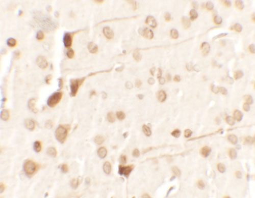
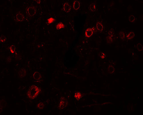
| WB, IHC-P, IF, E |
|---|---|
| Primary Accession | P13693 |
| Other Accession | NP_003286, 4507669 |
| Reactivity | Human, Mouse, Rat |
| Host | Rabbit |
| Clonality | Polyclonal |
| Isotype | IgG |
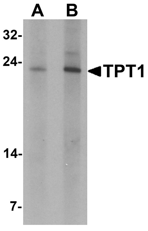
| Calculated MW | Predicted: 19 kDa |
| Application Notes | TPT1 antibody can be used for detection of TPT1 by Western blot at 0.5 - 1 µg/mL. |
| Gene ID | 7178 |
|---|---|
| Target/Specificity | TPT1; |
| Reconstitution & Storage | TPT1 antibody can be stored at 4℃ for three months and -20℃, stable for up to one year. |
| Precautions | TPT1 Antibody is for research use only and not for use in diagnostic or therapeutic procedures. |
| Name | TPT1 |
|---|---|
| Function | Involved in calcium binding and microtubule stabilization (PubMed:12167714, PubMed:15162379, PubMed:15958728). Acts as a negative regulator of TSC22D1-mediated apoptosis, via interaction with and destabilization of TSC22D1 protein (PubMed:18325344). |
| Cellular Location | Cytoplasm. |
| Tissue Location | Found in several healthy and tumoral cells including erythrocytes, hepatocytes, macrophages, platelets, keratinocytes, erythroleukemia cells, gliomas, melanomas, hepatoblastomas, and lymphomas. It cannot be detected in kidney and renal cell carcinoma (RCC). Expressed in placenta and prostate |

Thousands of laboratories across the world have published research that depended on the performance of antibodies from Abcepta to advance their research. Check out links to articles that cite our products in major peer-reviewed journals, organized by research category.
info@abcepta.com, and receive a free "I Love Antibodies" mug.

Provided below are standard protocols that you may find useful for product applications.
Background
TPT1 Antibody: TPT1 (translationally controlled tumor protein 1) is a 23 kDa member of the TCTP family of calcium binding proteins. TPT1 is localized in the cytoplasm and widely expressed and serves as a transcriptional activator, calcium transporter, histamine inducer and antiapoptotic caspase 3 inhibitor. TPT1 is a cytokine-like molecule that causes the release of histamine, IL-4 and IL-13 from basophils as well as the secretion of IL-8 and a calcium response in eosinophils. TPT1 plays a pivotal role in allergic diseases and due to its wide distribution in brain, is thought to be involved in neurodegenerative disorders, such as Alzheimer’s disease and Down syndrome.
References
MacDonald SM, Rafnar T, Langdon J, et al. Molecular identification of an IgE-dependent histamine-releasing factor. Science 1995; 269:688-90.
MacDonald SM. Human recombinant histamine-releasing factor. Int. Arch. Allergy Immunol. 1997; 113:187-9.
Sanchez JC, Schaller D, Ravier F, et al. Translationally controlled tumor protein: a protein identified in several nontumoral cells including erythrocytes. Electrophoresis 1997; 18:150-5.
Kuna P and Kaplan AP. Relationship of histamine-releasing factors and histamine-releasing inhibitory factors to chemokine group of cytokine. Allergy Asthma Proc. 1996; 17:5-11.
If you have used an Abcepta product and would like to share how it has performed, please click on the "Submit Review" button and provide the requested information. Our staff will examine and post your review and contact you if needed.
If you have any additional inquiries please email technical services at tech@abcepta.com.
Ordering Information
Other Products
Shipping Information
