


 Foundational characteristics of cancer include proliferation, angiogenesis, migration, evasion of apoptosis, and cellular immortality. Find key markers for these cellular processes and antibodies to detect them.
 The SUMOplot™ Analysis Program predicts and scores sumoylation sites in your protein. SUMOylation is a post-translational modification involved in various cellular processes, such as nuclear-cytosolic transport, transcriptional regulation, apoptosis, protein stability, response to stress, and progression through the cell cycle.
 The Autophagy Receptor Motif Plotter predicts and scores autophagy receptor binding sites in your protein. Identifying proteins connected to this pathway is critical to understanding the role of autophagy in physiological as well as pathological processes such as development, differentiation, neurodegenerative diseases, stress, infection, and cancer.
IL-28B Antibody
- SPECIFICATION
- CITATIONS
- PROTOCOLS
- BACKGROUND

Application
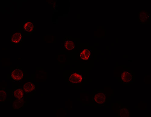
| WB, IF, E |
|---|---|
| Primary Accession | Q8IZI9 |
| Other Accession | NP_742151, 28144901 |
| Reactivity | Human |
| Host | Rabbit |
| Clonality | Polyclonal |
| Isotype | IgG |
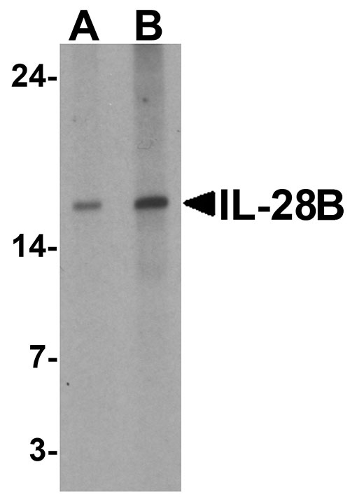
| Calculated MW | Predicted: 20 kDa Observed: 16kDa |
| Application Notes | IL-28B antibody can be used for detection of IL-28B by Western blot at 1 - 2 µg/ml. |
| Gene ID | 282617 |
|---|---|
| Target/Specificity | IFNL3; IL-28B antibody is human specific. IL-28B antibody may cross-react with IL-28A and IL-29. |
| Reconstitution & Storage | IL-28B antibody can be stored at 4℃ for three months and -20℃, stable for up to one year. |
| Precautions | IL-28B Antibody is for research use only and not for use in diagnostic or therapeutic procedures. |
| Name | IFNL3 |
|---|---|
| Synonyms | IL28B, IL28C, ZCYTO22 |
| Function | Cytokine with antiviral, antitumour and immunomodulatory activities. Plays a critical role in the antiviral host defense, predominantly in the epithelial tissues. Acts as a ligand for the heterodimeric class II cytokine receptor composed of IL10RB and IFNLR1, and receptor engagement leads to the activation of the JAK/STAT signaling pathway resulting in the expression of IFN-stimulated genes (ISG), which mediate the antiviral state. Has a restricted receptor distribution and therefore restricted targets: is primarily active in epithelial cells and this cell type-selective action is because of the epithelial cell-specific expression of its receptor IFNLR1. Seems not to be essential for early virus-activated host defense in vaginal infection, but plays an important role in Toll-like receptor (TLR)- induced antiviral defense. Plays a significant role in the antiviral immune defense in the intestinal epithelium. Exerts an immunomodulatory effect by up-regulating MHC class I antigen expression. |
| Cellular Location | Secreted. |

Thousands of laboratories across the world have published research that depended on the performance of antibodies from Abcepta to advance their research. Check out links to articles that cite our products in major peer-reviewed journals, organized by research category.
info@abcepta.com, and receive a free "I Love Antibodies" mug.

Provided below are standard protocols that you may find useful for product applications.
Background
Interleukin 28 (IL-28B), also known as interferon lambda 3, is cytokine distantly related to type I interferons and the IL-10 family (1). The IL-28B gene and the closely related cytokine genes IL-28A and IL-29 form a cytokine gene cluster on a chromosomal region mapped to 19q13. Expression of the cytokines encoded by the three genes can be induced by viral infection and can induce anti-viral activity (2,3). All three cytokines have been shown to interact with a heterodimeric class II cytokine receptor that consists of interleukin 10 receptor-beta (IL10RB) and interleukin 28 receptor- alpha (IL28RA) (1).
References
Sheppard P, Kindsvogel W, Xu W, et al. IL-28, IL-29 and their class II cytokine receptor IL-28R. Nat. Immunol. 2003; 4:63-8.
Osterlund PI, Pietila TE, Veckman V, et al. IFN regulatory factor family members differentially regulate the expression of type III IFN (IFN-lambda) genes. J. Immunol. 2007; 179:3434-42.
Meager A, Visvalingam K, Dilger P, et al. Biological activity of interleukins-28 and -29: comparison with type I interferons. Cytokine 2005; 31:109-18.
If you have used an Abcepta product and would like to share how it has performed, please click on the "Submit Review" button and provide the requested information. Our staff will examine and post your review and contact you if needed.
If you have any additional inquiries please email technical services at tech@abcepta.com.
Ordering Information
Other Products
Shipping Information
