


 Foundational characteristics of cancer include proliferation, angiogenesis, migration, evasion of apoptosis, and cellular immortality. Find key markers for these cellular processes and antibodies to detect them.
 The SUMOplot™ Analysis Program predicts and scores sumoylation sites in your protein. SUMOylation is a post-translational modification involved in various cellular processes, such as nuclear-cytosolic transport, transcriptional regulation, apoptosis, protein stability, response to stress, and progression through the cell cycle.
 The Autophagy Receptor Motif Plotter predicts and scores autophagy receptor binding sites in your protein. Identifying proteins connected to this pathway is critical to understanding the role of autophagy in physiological as well as pathological processes such as development, differentiation, neurodegenerative diseases, stress, infection, and cancer.
HSP70 Antibody
HSP70 Antibody, Clone 1H11
- SPECIFICATION
- CITATIONS
- PROTOCOLS
- BACKGROUND

Application
| WB, ICC, FC |
|---|---|
| Primary Accession | P0DMV8 |
| Other Accession | NP_005336.3 |
| Host | Mouse |
| Isotype | IgG1 |
| Reactivity | Human, Mouse, Rat, Pig, Bovine |
| Clonality | Monoclonal |
| Description | Mouse Anti-Human Cell Membrane HSP70 Monoclonal IgG1 |
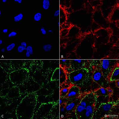
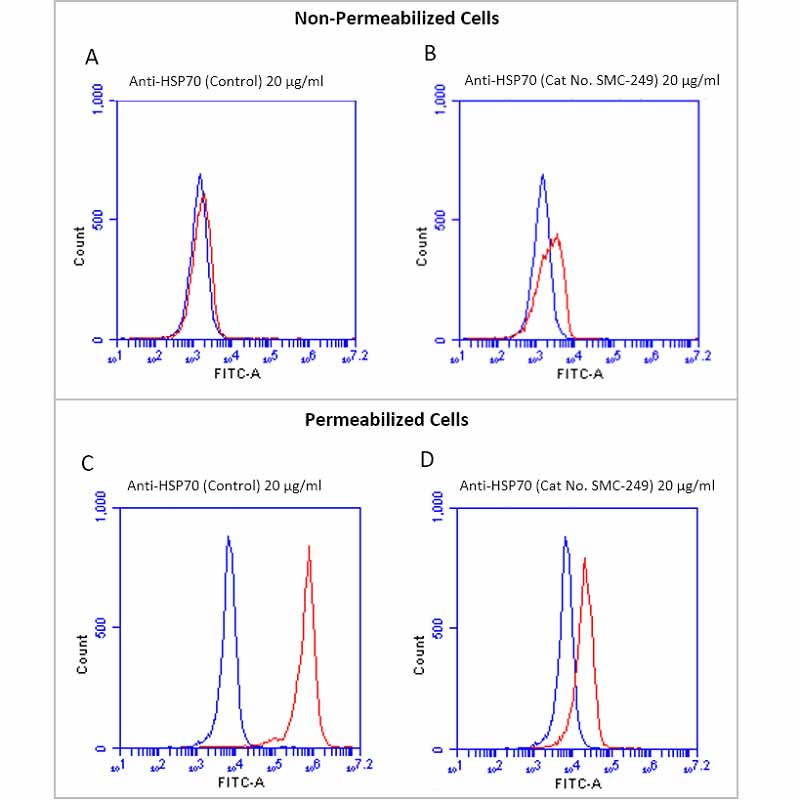
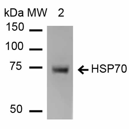
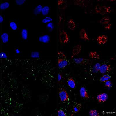
| Target/Specificity | Binds to cell surface HSP70 without permeabilization of cell membrane in tumor cell lines. Does not bind to intracellular HSP70 at low concentrations. Detects a ~70 kDa band in Western Blot. |
| Other Names | HSP70 1 Antibody, HSP70 2 Antibody, HSP70.1 Antibody, HSP72 Antibody, HSPA1 Antibody, HSPA1A Antibody, HSPA1B Antibody |
| Clone Names | 1H11 |
| Immunogen | Human native HSP70 protein |
| Purification | Protein G Purified |
| Storage | -20ºC |
| Storage Buffer | PBS pH 7.4, 50% glycerol, 0.9% Sodium Azide |
| Shipping Temperature | Blue Ice or 4ºC |
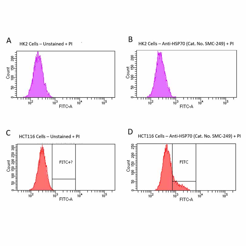
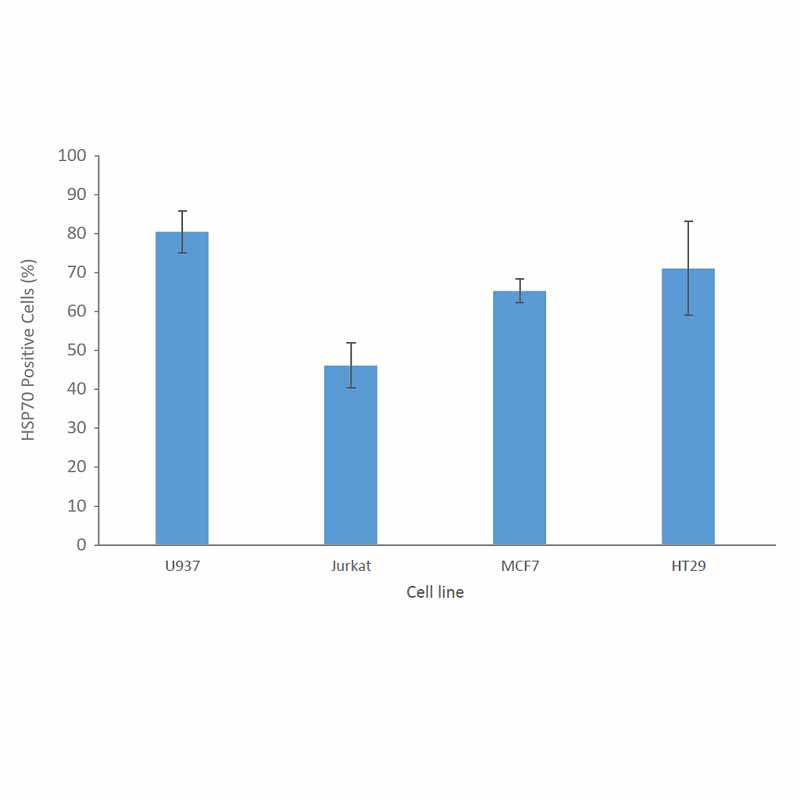
| Certificate of Analysis | A 1:250 dilution of SMC-249 was sufficient for detection of cell surface HSP70 in HCT116 cells using Fluorescence-activated cell sorting, applied in flow cytometry, with FITC as the fluorescent probe. |
| Cellular Localization | Cell membrane |

Thousands of laboratories across the world have published research that depended on the performance of antibodies from Abcepta to advance their research. Check out links to articles that cite our products in major peer-reviewed journals, organized by research category.
info@abcepta.com, and receive a free "I Love Antibodies" mug.

Provided below are standard protocols that you may find useful for product applications.
Background
HSP70 is a highly conserved protein that is ubiquitously expressed. It can be found in chloroplasts, eukaryotic cytosol, endoplasmic reticulum, and mitochondria, but also embedded in the cell membrane and in the extracellular space (1,2,3,4). Even though HSP70 is one of the most studied heat shock proteins, the export mechanism and method of membrane insertion are not fully understood. The majority of the proteins in the heat shock family lack a consensus signal for secretion via the ER-Golgi pathway (5). Researchers have found that HSP70 may be released from cells via exosomes or secretory vesicles (6). Although HSP70 is ubiquitously expressed, there is not much information about its presence on cell surface. The finding that HSP70 is localized on the cell surface of cancer cells but not normal cells suggests a conformational change of HSP70 in the lower pH environment characteristic of cancer cells (7). The presence of cell membrane embedded HSP70 has been found to increase the stability of cancer cells, thereby protecting tumors from environmental stress (8, 9). Anti-HSP70 antibody, clone 1H11 (Catalog No. SMC-249) is unique from other commercially available antibodies in that it can bind to the extracellular region of the cell membrane embedded HSP70 protein, allowing researchers to differentiate between membrane bound and intracellular HSP70 across cancer cells types.
References
1. Gribaldo, S. et al. (1999) J. Bacteriol. 181, 434-443.
2. Sharma, D. & Masison, D.C. (2009) Protein Pept. Lett. 16, 571-581.
3. Sharma, D. et al. (2009) PLoS. ONE.4, e6644.
4. Kampinga, H.H. & Craig, E.A. (2010) Nat. Rev. Mol. Cell Biol. 11, 579-592.
5. Nickel, W. & Seedorf, M. (2008) Annu. Rev. Cell Dev. Biol. 24, 287-308.
6. Multhoff, G. & Hightower, L.E. (1996) Cell Stress. Chaperones. 1, 167-176.
7. De Maio, A. (2011) Cell Stress. Chaperones. 16, 235-249.
8. Horvath, I. & Vigh, L. (2010) Nature. 463, 436-438.
9. Horvath, I., Multhoff, G., Sonnleitner, A., & Vigh, L. (2008) Biochim. Biophys. Acta 1778, 1653-1664.
If you have used an Abcepta product and would like to share how it has performed, please click on the "Submit Review" button and provide the requested information. Our staff will examine and post your review and contact you if needed.
If you have any additional inquiries please email technical services at tech@abcepta.com.
Ordering Information
Other Products
Shipping Information
