


 Foundational characteristics of cancer include proliferation, angiogenesis, migration, evasion of apoptosis, and cellular immortality. Find key markers for these cellular processes and antibodies to detect them.
 The SUMOplot™ Analysis Program predicts and scores sumoylation sites in your protein. SUMOylation is a post-translational modification involved in various cellular processes, such as nuclear-cytosolic transport, transcriptional regulation, apoptosis, protein stability, response to stress, and progression through the cell cycle.
 The Autophagy Receptor Motif Plotter predicts and scores autophagy receptor binding sites in your protein. Identifying proteins connected to this pathway is critical to understanding the role of autophagy in physiological as well as pathological processes such as development, differentiation, neurodegenerative diseases, stress, infection, and cancer.
Anti-HUMAN HAPTOGLOBIN (RABBIT) Antibody
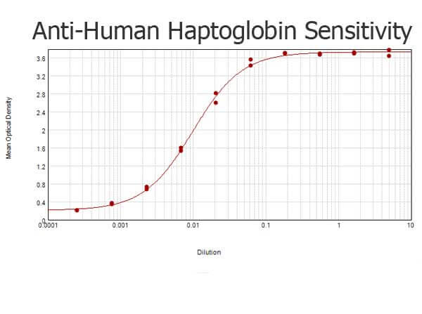
Human Haptoglobin Antibody
- SPECIFICATION
- CITATIONS
- PROTOCOLS
- BACKGROUND

| Host | Rabbit |
|---|---|
| Conjugate | Unconjugated |
| Target Species | Human |
| Reactivity | Human |
| Clonality | Polyclonal |
Application
| WB, IHC, E, I, LCI |
| Application Note | Human Haptoglobin Antibody is tested by ELISA and is suitable for western blotting, ELISA and IHC. Researchers should determine optimal titers for applications that are not stated below. |
| Physical State | Liquid (sterile filtered) |
| Buffer | 0.02 M Potassium Phosphate, 0.15 M Sodium Chloride, pH 7.2 |
| Immunogen | Human Haptoglobin Antibody was produced by repeated immunizations with Human Haptoglobin protein. |
| Preservative | 0.01% (w/v) Sodium Azide |
| Gene ID | 3240 |
|---|---|
| Other Names | 3240 |
| Purity | Human Haptoglobin Antibody was prepared from monospecific antiserum by immunoaffinity chromatography using antigen resins. Assay by immunoelectrophoresis resulted in a single precipitin arc against anti-Rabbit Serum. Analysis by SDS-PAGE was used to determine that this preparation is substantially free of aggregates and shows a banding pattern consistent with purified Rabbit IgG. |
| Storage Condition | Store vial at -20° C prior to opening. Aliquot contents and freeze at -20° C or below for extended storage. Avoid cycles of freezing and thawing. Centrifuge product if not completely clear after standing at room temperature. This product is stable for several weeks at 4° C as an undiluted liquid. Dilute only prior to immediate use. |
| Precautions Note | This product is for research use only and is not intended for therapeutic or diagnostic applications. |
| Name | HP |
|---|---|
| Function | As a result of hemolysis, hemoglobin is found to accumulate in the kidney and is secreted in the urine. Haptoglobin captures, and combines with free plasma hemoglobin to allow hepatic recycling of heme iron and to prevent kidney damage. Haptoglobin also acts as an antioxidant, has antibacterial activity, and plays a role in modulating many aspects of the acute phase response. Hemoglobin/haptoglobin complexes are rapidly cleared by the macrophage CD163 scavenger receptor expressed on the surface of liver Kupfer cells through an endocytic lysosomal degradation pathway. |
| Cellular Location | Secreted. |
| Tissue Location | Expressed by the liver and secreted in plasma. |

Thousands of laboratories across the world have published research that depended on the performance of antibodies from Abcepta to advance their research. Check out links to articles that cite our products in major peer-reviewed journals, organized by research category.
info@abcepta.com, and receive a free "I Love Antibodies" mug.

Provided below are standard protocols that you may find useful for product applications.
Background
Anti-Human Haptoglobin Antibody detects Haptoglobin. Haptoglobin combined with free plasma hemoglobin prevents loss of iron through the kidneys and protects the kidneys from damage by hemoglobin, while making the hemoglobin accessible to degradative enzymes. Haptoglobin is expressed by the liver and secreted in plasma. Anti-Haptoglobin Antibody is ideal for investigators involved in Cell Signaling, Immunology and Signal Transduction research.
If you have used an Abcepta product and would like to share how it has performed, please click on the "Submit Review" button and provide the requested information. Our staff will examine and post your review and contact you if needed.
If you have any additional inquiries please email technical services at tech@abcepta.com.
Ordering Information
Other Products
Shipping Information
