


 Foundational characteristics of cancer include proliferation, angiogenesis, migration, evasion of apoptosis, and cellular immortality. Find key markers for these cellular processes and antibodies to detect them.
 The SUMOplot™ Analysis Program predicts and scores sumoylation sites in your protein. SUMOylation is a post-translational modification involved in various cellular processes, such as nuclear-cytosolic transport, transcriptional regulation, apoptosis, protein stability, response to stress, and progression through the cell cycle.
 The Autophagy Receptor Motif Plotter predicts and scores autophagy receptor binding sites in your protein. Identifying proteins connected to this pathway is critical to understanding the role of autophagy in physiological as well as pathological processes such as development, differentiation, neurodegenerative diseases, stress, infection, and cancer.
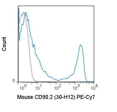
PE-Cy7 Anti-Mouse CD90.2 (30-H12) Antibody
- SPECIFICATION
- CITATIONS
- PROTOCOLS
- BACKGROUND

Application
| FC |
|---|---|
| Isotype | Rat IgG2b |
| Concentration | 0.2 mg/mL |
| Reactivity | Mouse |
| Formulation | 10 mM NaH2PO4, 150 mM NaCl, 0.09% NaN3, 0.1% gelatin, pH7.2 |
| Host | Rat |
| Gene ID | 21838 |
|---|---|
| Gene Name | Thy1 |
| Alternative Name(s) | Thy1.2 |
| Format | PE-Cy7 |
| Preparation | This monoclonal antibody was purified from tissue culture supernatant via affinity chromatography. The purified antibody was conjugated under optimal conditions, with unreacted dye removed from the preparation. It is recommended to store the product undiluted at 4°C, and protected from prolonged exposure to light. Do not freeze. |
| Application Notes | pe-cy7-anti-mouse-thy-1-2-30-h12.html |
| Storage Conditions | 2-8°C protected from light |
Ledbetter JA and Herzenberg LA. 1979. Immunol. Rev. 47:63-90. Borrello MA and Phipps RP. 1996. Cell Immunol. 173:198-206. (flow cytometry)
Seaman WE, Wofsy D, Greenspan JS, and Ledbetter JA. 1983. J. Immunol. 130: 1713-8. (depletion)
Provided below are standard protocols that you may find useful for product applications.
Background
The 30-H12 antibody reacts with mouse CD90.2 (Thy1.2). CD90.2 is a strain-specific allelic form of the GPI-linked membrane associated protein CD90 and is involved in adhesion and signal transduction. CD90.2 is expressed on thymocytes, mature T cells and neurons in mouse strains that express the CD90.2 allele (BALB/c, CBA, C3H, C57BL/6, SJL and others). 30-H12 does not react with the CD90.1 allele expressed in mouse strains such as PL and AKR.

If you have used an Abcepta product and would like to share how it has performed, please click on the "Submit Review" button and provide the requested information. Our staff will examine and post your review and contact you if needed.
If you have any additional inquiries please email technical services at tech@abcepta.com.
Ordering Information
Other Products
Shipping Information
