


 Foundational characteristics of cancer include proliferation, angiogenesis, migration, evasion of apoptosis, and cellular immortality. Find key markers for these cellular processes and antibodies to detect them.
 The SUMOplot™ Analysis Program predicts and scores sumoylation sites in your protein. SUMOylation is a post-translational modification involved in various cellular processes, such as nuclear-cytosolic transport, transcriptional regulation, apoptosis, protein stability, response to stress, and progression through the cell cycle.
 The Autophagy Receptor Motif Plotter predicts and scores autophagy receptor binding sites in your protein. Identifying proteins connected to this pathway is critical to understanding the role of autophagy in physiological as well as pathological processes such as development, differentiation, neurodegenerative diseases, stress, infection, and cancer.
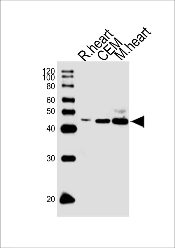
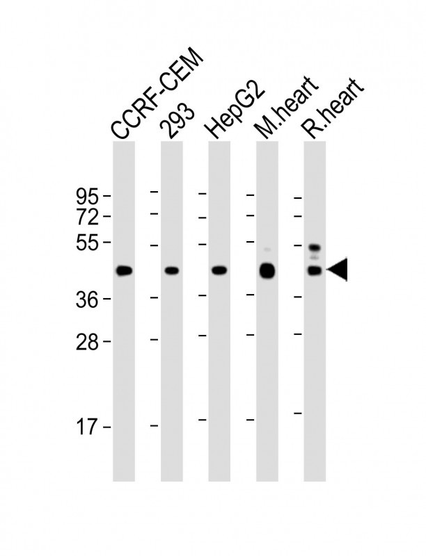
VEGFC Antibody
Mouse Monoclonal Antibody (Mab)
- SPECIFICATION
- CITATIONS
- PROTOCOLS
- BACKGROUND

Application
| WB |
|---|---|
| Primary Accession | P49767 |
| Other Accession | NP_005420.1 |
| Reactivity | Human, Mouse, Rat |
| Host | Mouse |
| Clonality | Monoclonal |
| Calculated MW | H=47;M=46;Rat=46 KDa |
| Isotype | IgG1,K |
| Antigen Source | HUMAN |
| Gene ID | 7424 |
|---|---|
| Antigen Region | 1-270 aa |
| Other Names | VEGFC; Vascular endothelial growth factor C; Flt4 ligand; Vascular endothelial growth factor-related protein |
| Dilution | WB~~1:2000 |
| Target/Specificity | This VEGFC monoclonal antibody is generated from mouse immunized with VEGFC recombinant protein. |
| Format | Purified monoclonal antibody supplied in PBS with 0.09% (W/V) sodium azide. This antibody is purified through a protein G column, followed by dialysis against PBS. |
| Storage | Maintain refrigerated at 2-8°C for up to 2 weeks. For long term storage store at -20°C in small aliquots to prevent freeze-thaw cycles. |
| Precautions | VEGFC Antibody is for research use only and not for use in diagnostic or therapeutic procedures. |
| Name | VEGFC |
|---|---|
| Function | Growth factor active in angiogenesis, and endothelial cell growth, stimulating their proliferation and migration and also has effects on the permeability of blood vessels. May function in angiogenesis of the venous and lymphatic vascular systems during embryogenesis, and also in the maintenance of differentiated lymphatic endothelium in adults. Binds and activates KDR/VEGFR2 and FLT4/VEGFR3 receptors. |
| Cellular Location | Secreted. |
| Tissue Location | Expressed in the spleen (PubMed:8700872, PubMed:9247316). Expressed in the lymph node, thymus, appendix and bone marrow (PubMed:9247316). Expressed in the heart, placenta, skeletal muscle, ovary and small intestine (PubMed:8617204, PubMed:8700872) Expressed in the prostate, testis and colon (PubMed:8700872) |

Thousands of laboratories across the world have published research that depended on the performance of antibodies from Abcepta to advance their research. Check out links to articles that cite our products in major peer-reviewed journals, organized by research category.
info@abcepta.com, and receive a free "I Love Antibodies" mug.

Provided below are standard protocols that you may find useful for product applications.
Background
The protein encoded by this gene is a member of the platelet-derived growth factor/vascular endothelial growth factor (PDGF/VEGF) family, is active in angiogenesis and endothelial cell growth, and can also affect the permeability of blood vessels. This secreted protein undergoes a complex proteolytic maturation, generating multiple processed forms which bind and activate VEGFR-3 receptors. Only the fully processed form can bind and activate VEGFR-2 receptors. This protein is structurally and functionally similar to vascular endothelial growth factor D. [provided by RefSeq].
References
Chen, X., et al. Cancer Sci. 101(11):2384-2390(2010)
Romero, R., et al. Am. J. Obstet. Gynecol. 203 (4), 361 (2010) :
Bailey, S.D., et al. Diabetes Care 33(10):2250-2253(2010)
Deguchi, K., et al. Anticancer Res. 30(6):2361-2366(2010)
Johnatty, S.E., et al. PLoS Genet. 6 (7), E1001016 (2010) :
If you have used an Abcepta product and would like to share how it has performed, please click on the "Submit Review" button and provide the requested information. Our staff will examine and post your review and contact you if needed.
If you have any additional inquiries please email technical services at tech@abcepta.com.

Ordering Information
Other Products
Shipping Information
