


 Foundational characteristics of cancer include proliferation, angiogenesis, migration, evasion of apoptosis, and cellular immortality. Find key markers for these cellular processes and antibodies to detect them.
 The SUMOplot™ Analysis Program predicts and scores sumoylation sites in your protein. SUMOylation is a post-translational modification involved in various cellular processes, such as nuclear-cytosolic transport, transcriptional regulation, apoptosis, protein stability, response to stress, and progression through the cell cycle.
 The Autophagy Receptor Motif Plotter predicts and scores autophagy receptor binding sites in your protein. Identifying proteins connected to this pathway is critical to understanding the role of autophagy in physiological as well as pathological processes such as development, differentiation, neurodegenerative diseases, stress, infection, and cancer.
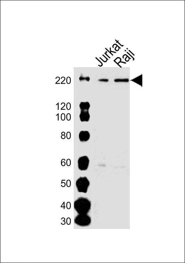
DOCK2 Antibody (C-term)
Purified Rabbit Polyclonal Antibody (Pab)
- SPECIFICATION
- CITATIONS
- PROTOCOLS
- BACKGROUND

Application
| WB |
|---|---|
| Primary Accession | Q92608 |
| Reactivity | Human |
| Predicted | Mouse |
| Host | Rabbit |
| Clonality | Polyclonal |
| Calculated MW | H=212;M=212 KDa |
| Isotype | Rabbit IgG |
| Antigen Source | HUMAN |
| Gene ID | 1794 |
|---|---|
| Antigen Region | 1812-1846 aa |
| Other Names | Dedicator of cytokinesis protein 2, DOCK2, KIAA0209 |
| Dilution | WB~~1:1000 |
| Target/Specificity | This DOCK2 antibody is generated from a rabbit immunized with a KLH conjugated synthetic peptide between 1812-1846 amino acids from the C-terminal region of human DOCK2. |
| Format | Purified polyclonal antibody supplied in PBS with 0.09% (W/V) sodium azide. This antibody is purified through a protein A column, followed by peptide affinity purification. |
| Storage | Maintain refrigerated at 2-8°C for up to 2 weeks. For long term storage store at -20°C in small aliquots to prevent freeze-thaw cycles. |
| Precautions | DOCK2 Antibody (C-term) is for research use only and not for use in diagnostic or therapeutic procedures. |
| Name | DOCK2 |
|---|---|
| Synonyms | KIAA0209 |
| Function | Involved in cytoskeletal rearrangements required for lymphocyte migration in response of chemokines. Activates RAC1 and RAC2, but not CDC42, by functioning as a guanine nucleotide exchange factor (GEF), which exchanges bound GDP for free GTP. May also participate in IL2 transcriptional activation via the activation of RAC2. |
| Cellular Location | Endomembrane system; Peripheral membrane protein. Cytoplasm, cytoskeleton. Note=Colocalizes with F-actin |
| Tissue Location | Specifically expressed in hematopoietic cells. Highly expressed in peripheral blood leukocytes, and expressed at intermediate level in thymus and spleen. Expressed at very low level in the small intestine and colon. |

Thousands of laboratories across the world have published research that depended on the performance of antibodies from Abcepta to advance their research. Check out links to articles that cite our products in major peer-reviewed journals, organized by research category.
info@abcepta.com, and receive a free "I Love Antibodies" mug.

Provided below are standard protocols that you may find useful for product applications.
Background
Involved in cytoskeletal rearrangements required for lymphocyte migration in response of chemokines. Activates RAC1 and RAC2, but not CDC42, by functioning as a guanine nucleotide exchange factor (GEF), which exchanges bound GDP for free GTP. May also participate in IL2 transcriptional activation via the activation of RAC2.
References
Nagase T.,et al.DNA Res. 3:321-329(1996).
Nishihara H.,et al.Biochim. Biophys. Acta 1452:179-187(1999).
Nishihara H.,et al.Blood 100:3968-3974(2002).
Nishihara H.,et al.Biochem. Biophys. Res. Commun. 296:716-720(2002).
Cote J.-F.,et al.J. Cell Sci. 115:4901-4913(2002).
If you have used an Abcepta product and would like to share how it has performed, please click on the "Submit Review" button and provide the requested information. Our staff will examine and post your review and contact you if needed.
If you have any additional inquiries please email technical services at tech@abcepta.com.
Ordering Information
Other Products
Shipping Information
