


 Foundational characteristics of cancer include proliferation, angiogenesis, migration, evasion of apoptosis, and cellular immortality. Find key markers for these cellular processes and antibodies to detect them.
 The SUMOplot™ Analysis Program predicts and scores sumoylation sites in your protein. SUMOylation is a post-translational modification involved in various cellular processes, such as nuclear-cytosolic transport, transcriptional regulation, apoptosis, protein stability, response to stress, and progression through the cell cycle.
 The Autophagy Receptor Motif Plotter predicts and scores autophagy receptor binding sites in your protein. Identifying proteins connected to this pathway is critical to understanding the role of autophagy in physiological as well as pathological processes such as development, differentiation, neurodegenerative diseases, stress, infection, and cancer.
Anti-P2X6 Antibody
- SPECIFICATION
- CITATIONS
- PROTOCOLS
- BACKGROUND

Application
| WB |
|---|---|
| Primary Accession | O15547 |
| Host | Rabbit |
| Reactivity | Human |
| Clonality | Polyclonal |
| Format | Lyophilized |
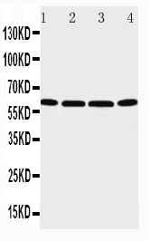
| Description | Rabbit IgG polyclonal antibody for P2X purinoceptor 6(P2RX6) detection. Tested with WB in Human. |
| Reconstitution | Add 0.2ml of distilled water will yield a concentration of 500ug/ml. |
| Gene ID | 9127 |
|---|---|
| Other Names | P2X purinoceptor 6, P2X6, ATP receptor, P2XM, Purinergic receptor, Purinergic receptor P2X-like 1, P2RX6, P2RXL1, P2X6 |
| Calculated MW | 48829 MW KDa |
| Application Details | Western blot, 0.1-0.5 µg/ml, Human |
| Subcellular Localization | Membrane; Multi-pass membrane protein. |
| Tissue Specificity | Expressed predominantly in skeletal muscle. |
| Protein Name | P2X purinoceptor 6 |
| Contents | Each vial contains 5mg BSA, 0.9mg NaCl, 0.2mg Na2HPO4, 0.05mg Thimerosal, 0.05mg NaN3. |
| Immunogen | A synthetic peptide corresponding to a sequence at the C-terminus of human P2X6(387-406aa VWRELALASQARLAECLRRS). |
| Purification | Immunogen affinity purified. |
| Cross Reactivity | No cross reactivity with other proteins |
| Storage | At -20˚C for one year. After r˚Constitution, at 4˚C for one month. It˚Can also be aliquotted and stored frozen at -20˚C for a longer time.Avoid repeated freezing and thawing. |
| Sequence Similarities | Belongs to the P2X receptor family. |
| Name | P2RX6 |
|---|---|
| Synonyms | P2RXL1, P2X6 |
| Function | May act as a modulatory subunit rather than a functional channel. Unlike other P2XRs members, P2RX6 does not seem to form functional homotrimers (PubMed:22378790). P2RX6 requires the presence of P2RX4 or P2RX2 to shuttle it to the plasma membrane where it may form functional heterotrimeric receptors at the plasma membrane (PubMed:22378790). P2RX6 can be translocated to the nucleus and functions as a nuclear regulator of post-transcriptional modifications in neurons (By similarity). |
| Cellular Location | Cell membrane {ECO:0000250|UniProtKB:P51579}; Multi-pass membrane protein. Endoplasmic reticulum {ECO:0000250|UniProtKB:P51579}. Nucleus {ECO:0000250|UniProtKB:O54803} Nucleus inner membrane {ECO:0000250|UniProtKB:O54803}; Multi-pass membrane protein. Note=Heteromerization of P2RX6 subunits with either P2RX2 or P2RX4 subunits guides the P2RX6 subunit to the plasma membrane (By similarity). Monomers remain anchored to the endoplasmic reticulum (ER) membrane by the hydrophobic N-terminal end (By similarity). Mainly expressed in the cell body of the hippocampal neurons (By similarity). {ECO:0000250|UniProtKB:O54803, ECO:0000250|UniProtKB:P51579} |
| Tissue Location | Expressed predominantly in skeletal muscle. |

Thousands of laboratories across the world have published research that depended on the performance of antibodies from Abcepta to advance their research. Check out links to articles that cite our products in major peer-reviewed journals, organized by research category.
info@abcepta.com, and receive a free "I Love Antibodies" mug.

Provided below are standard protocols that you may find useful for product applications.
Background
Purinergic receptor P2X-Like 1, also known as P2X6, is a protein that in humans is encoded by the P2RX6 gene. The encoded protein is associated with VE-cadherin at the adherens junctions of human umbilical vein endothelial cells. This gene belongs to the family of P2X receptors. P2RXL1 gene was mapped to chromosome 22q11 by fluorescence in situ hybridization. This gene is a receptor for ATP that acts as a ligand-gated ion channel. It can mediate rapid and selective permeability to cations.
If you have used an Abcepta product and would like to share how it has performed, please click on the "Submit Review" button and provide the requested information. Our staff will examine and post your review and contact you if needed.
If you have any additional inquiries please email technical services at tech@abcepta.com.
Ordering Information
Other Products
Shipping Information
