


 Foundational characteristics of cancer include proliferation, angiogenesis, migration, evasion of apoptosis, and cellular immortality. Find key markers for these cellular processes and antibodies to detect them.
 The SUMOplot™ Analysis Program predicts and scores sumoylation sites in your protein. SUMOylation is a post-translational modification involved in various cellular processes, such as nuclear-cytosolic transport, transcriptional regulation, apoptosis, protein stability, response to stress, and progression through the cell cycle.
 The Autophagy Receptor Motif Plotter predicts and scores autophagy receptor binding sites in your protein. Identifying proteins connected to this pathway is critical to understanding the role of autophagy in physiological as well as pathological processes such as development, differentiation, neurodegenerative diseases, stress, infection, and cancer.
> home > Products > Primary Antibodies > Antibody Collections > KD-Validated Antibodies > KD-Validated Anti-ATP6V1A Rabbit Monoclonal Antibody
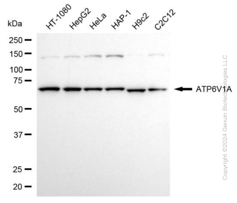
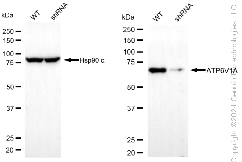
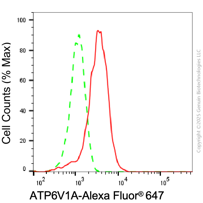
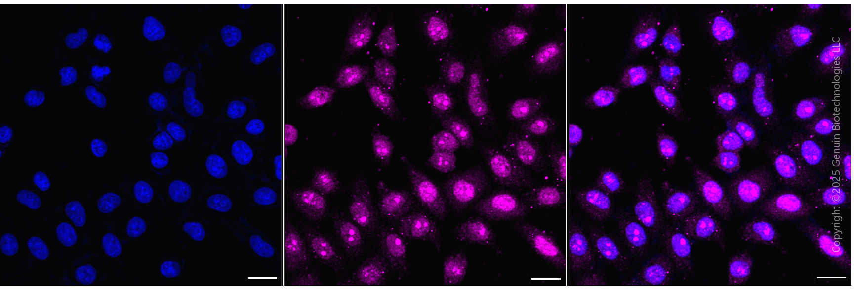
KD-Validated Anti-ATP6V1A Rabbit Monoclonal Antibody
Rabbit monoclonal antibody
- SPECIFICATION
- CITATIONS
- PROTOCOLS
- BACKGROUND

Application
| WB, FC, ICC |
|---|---|
| Primary Accession | P38606 |
| Reactivity | Rat, Human, Mouse |
| Clonality | Monoclonal |
| Isotype | Rabbit IgG |
| Clone Names | 24GB6940 |
| Calculated MW | Predicted, 68 kDa, observed, 68 kDa |
| Gene Name | ATP6V1A |
| Aliases | ATP6V1A; ATPase H+ Transporting V1 Subunit A; V-ATPase Subunit A; ATP6V1A1; ATP6A1; Vma1; VA68; VPP2; ATPase, H+ Transporting, Lysosomal 70kDa, V1 Subunit A; V-Type Proton ATPase (V-ATPase) Catalytic Subunit A; V-Type Proton ATPase Catalytic Subunit A; Vacuolar Proton Pump Subunit Alpha; ATPase, H+ Transporting, Lysosomal (Vacuolar Proton Pump), Alpha Polypeptide, 70kD, Isoform 1; H+-Transporting ATPase Chain A, Vacuolar (VA68 Type); ATPase, H+ Transporting, Lysosomal, Subunit A1; H(+)-Transporting Two-Sector ATPase, Subunit A; Vacuolar Proton Pump Alpha Subunit 1; Vacuolar ATPase Isoform VA68; V-ATPase 69 KDa Subunit 1; Vacuolar-Type H(+)-ATPase; V-ATPase 69 KDa Subunit; V-ATPase A Subunit 1; EC 3.6.3.14; EC 7.1.2.2; EC 3.6.3; ARCL2D; IECEE3; DEE93; HO68 |
| Immunogen | Recombinant protein of human ATP6V1A |
| Gene ID | 523 |
|---|---|
| Other Names | V-type proton ATPase catalytic subunit A, V-ATPase subunit A, 7.1.2.2, V-ATPase 69 kDa subunit, Vacuolar ATPase isoform VA68, Vacuolar proton pump subunit alpha, ATP6V1A, ATP6A1, ATP6V1A1, VPP2 |
| Name | ATP6V1A |
|---|---|
| Synonyms | ATP6A1, ATP6V1A1, VPP2 |
| Function | Catalytic subunit of the V1 complex of vacuolar(H+)-ATPase (V-ATPase), a multisubunit enzyme composed of a peripheral complex (V1) that hydrolyzes ATP and a membrane integral complex (V0) that translocates protons (PubMed:8463241). V-ATPase is responsible for acidifying and maintaining the pH of intracellular compartments and in some cell types, is targeted to the plasma membrane, where it is responsible for acidifying the extracellular environment (PubMed:32001091). In aerobic conditions, involved in intracellular iron homeostasis, thus triggering the activity of Fe(2+) prolyl hydroxylase (PHD) enzymes, and leading to HIF1A hydroxylation and subsequent proteasomal degradation (PubMed:28296633). May play a role in neurite development and synaptic connectivity (PubMed:29668857). |
| Cellular Location | Cytoplasm. Cytoplasm, cytosol {ECO:0000250|UniProtKB:P50516}. Cytoplasmic vesicle, secretory vesicle. Cytoplasmic vesicle, clathrin-coated vesicle membrane {ECO:0000250|UniProtKB:P31404}; Peripheral membrane protein. Lysosome {ECO:0000250|UniProtKB:P50516} Note=Co-localizes with WFS1 in the secretory granules in neuroblastoma cell lines. |
| Tissue Location | High expression in the skin. |
Research Areas
Citations (0)

Thousands of laboratories across the world have published research that depended on the performance of antibodies from Abcepta to advance their research. Check out links to articles that cite our products in major peer-reviewed journals, organized by research category.
Submit your citation using an Abcepta antibody to
info@abcepta.com, and receive a free "I Love Antibodies" mug.
info@abcepta.com, and receive a free "I Love Antibodies" mug.

Application Protocols
Provided below are standard protocols that you may find useful for product applications.
Abcepta welcomes feedback from its customers.
If you have used an Abcepta product and would like to share how it has performed, please click on the "Submit Review" button and provide the requested information. Our staff will examine and post your review and contact you if needed.
If you have any additional inquiries please email technical services at tech@abcepta.com.
$ 399.20

$ 149.00
Cat# AGI1982
Ordering Information
United States
AlbaniaAustraliaAustriaBelgiumBosnia & HerzegovinaBrazilBulgariaCanadaCentral AmericaChinaCroatiaCyprusCzech RepublicDenmarkEstoniaFinlandFranceGermanyGreeceHong KongHungaryIcelandIndiaIndonesiaIrelandIsraelItalyJapanLatviaLithuaniaLuxembourgMacedoniaMalaysiaMaltaMexicoNetherlandsNew ZealandNorwayPakistanPolandPortugalRomaniaSerbiaSingaporeSlovakiaSloveniaSouth AfricaSouth KoreaSpainSwedenSwitzerlandTaiwanTurkeyUnited KingdomUnited StatesVietnamWorldwideOthers
USA Headquarters
(888) 735-7227 / (858) 622-0099 or (858) 875-1900
Other Products
Shipping Information
Domestic orders (in stock items)
Shipped out the same day. Orders placed after 1 PM (PST) will ship out the next business day.
International orders
Contact your local distributors
