


 Foundational characteristics of cancer include proliferation, angiogenesis, migration, evasion of apoptosis, and cellular immortality. Find key markers for these cellular processes and antibodies to detect them.
 The SUMOplot™ Analysis Program predicts and scores sumoylation sites in your protein. SUMOylation is a post-translational modification involved in various cellular processes, such as nuclear-cytosolic transport, transcriptional regulation, apoptosis, protein stability, response to stress, and progression through the cell cycle.
 The Autophagy Receptor Motif Plotter predicts and scores autophagy receptor binding sites in your protein. Identifying proteins connected to this pathway is critical to understanding the role of autophagy in physiological as well as pathological processes such as development, differentiation, neurodegenerative diseases, stress, infection, and cancer.
> home > Products > Primary Antibodies > Antibody Collections > KD-Validated Antibodies > KD-Validated Anti-ATG5 Rabbit Polyclonal Antibody
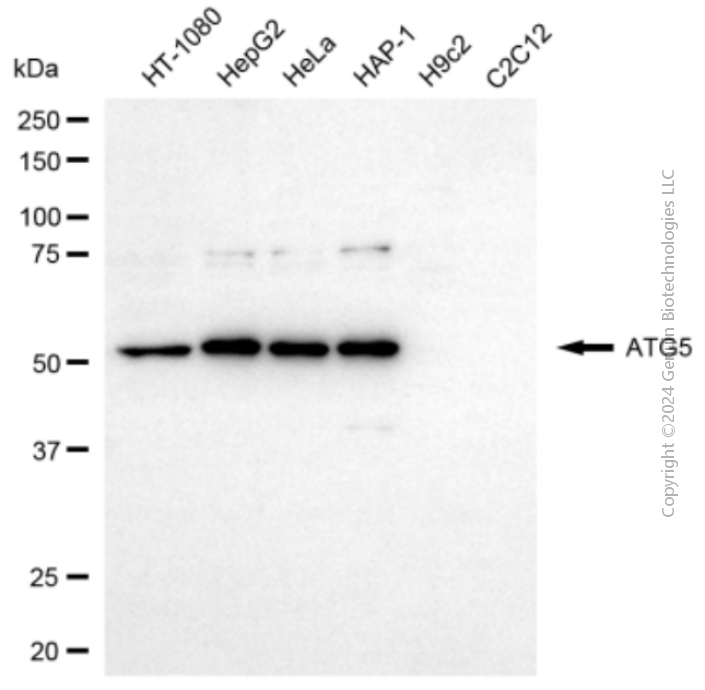
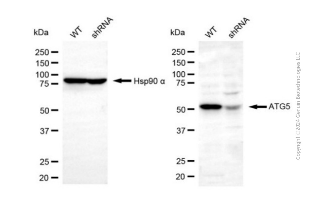
KD-Validated Anti-ATG5 Rabbit Polyclonal Antibody
Rabbit polyclonal antibody
- SPECIFICATION
- CITATIONS
- PROTOCOLS
- BACKGROUND

Application
| WB |
|---|---|
| Primary Accession | Q9H1Y0 |
| Reactivity | Human |
| Clonality | Polyclonal |
| Isotype | Rabbit IgG |
| Calculated MW | Predicted, 32 kDa, observed, 55 kDa |
| Gene Name | ATG5 |
| Aliases | ATG5; Autophagy Related 5; ASP; HAPG5; APG5L; APG5; Apoptosis-Specific Protein; Autophagy Protein 5; ATG5 Autophagy Related 5 Homolog (S. Cerevisiae); APG5 (Autophagy 5, S. Cerevisiae)-Like; APG5 Autophagy 5-Like (S. Cerevisiae); ATG5 Autophagy Related 5 Homolog; APG5 Autophagy 5-Like; APG5-LIKE; APG5-Like; SCAR25 |
| Immunogen | A synthesized peptide derived from human ATG5 |
| Gene ID | 9474 |
|---|---|
| Other Names | Autophagy protein 5, APG5-like, Apoptosis-specific protein, ATG5 (HGNC:589), APG5L, ASP |
| Name | ATG5 (HGNC:589) |
|---|---|
| Synonyms | APG5L, ASP |
| Function | Involved in autophagic vesicle formation. Conjugation with ATG12, through a ubiquitin-like conjugating system involving ATG7 as an E1-like activating enzyme and ATG10 as an E2-like conjugating enzyme, is essential for its function. The ATG12-ATG5 conjugate acts as an E3- like enzyme which is required for lipidation of ATG8 family proteins and their association to the vesicle membranes. Involved in mitochondrial quality control after oxidative damage, and in subsequent cellular longevity. Plays a critical role in multiple aspects of lymphocyte development and is essential for both B and T lymphocyte survival and proliferation. Required for optimal processing and presentation of antigens for MHC II. Involved in the maintenance of axon morphology and membrane structures, as well as in normal adipocyte differentiation. Promotes primary ciliogenesis through removal of OFD1 from centriolar satellites and degradation of IFT20 via the autophagic pathway. As part of the ATG8 conjugation system with ATG12 and ATG16L1, required for recruitment of LRRK2 to stressed lysosomes and induction of LRRK2 kinase activity in response to lysosomal stress (By similarity). |
| Cellular Location | Cytoplasm. Preautophagosomal structure membrane; Peripheral membrane protein. Note=Colocalizes with nonmuscle actin. The conjugate detaches from the membrane immediately before or after autophagosome formation is completed (By similarity). Also localizes to discrete punctae along the ciliary axoneme and to the base of the ciliary axoneme. Under starved conditions, the ATG12-ATG5-ATG16L1 complex is translocated to phagophores driven by RAB33B (PubMed:32960676). {ECO:0000250, ECO:0000269|PubMed:32960676} |
| Tissue Location | Ubiquitous. The mRNA is present at similar levels in viable and apoptotic cells, whereas the protein is dramatically highly expressed in apoptotic cells |
Research Areas
Citations (0)

Thousands of laboratories across the world have published research that depended on the performance of antibodies from Abcepta to advance their research. Check out links to articles that cite our products in major peer-reviewed journals, organized by research category.
Submit your citation using an Abcepta antibody to
info@abcepta.com, and receive a free "I Love Antibodies" mug.
info@abcepta.com, and receive a free "I Love Antibodies" mug.

Application Protocols
Provided below are standard protocols that you may find useful for product applications.
Abcepta welcomes feedback from its customers.
If you have used an Abcepta product and would like to share how it has performed, please click on the "Submit Review" button and provide the requested information. Our staff will examine and post your review and contact you if needed.
If you have any additional inquiries please email technical services at tech@abcepta.com.
$ 399.20

$ 149.00
Cat# AGI2155
Ordering Information
United States
AlbaniaAustraliaAustriaBelgiumBosnia & HerzegovinaBrazilBulgariaCanadaCentral AmericaChinaCroatiaCyprusCzech RepublicDenmarkEstoniaFinlandFranceGermanyGreeceHong KongHungaryIcelandIndiaIndonesiaIrelandIsraelItalyJapanLatviaLithuaniaLuxembourgMacedoniaMalaysiaMaltaMexicoNetherlandsNew ZealandNorwayPakistanPolandPortugalRomaniaSerbiaSingaporeSlovakiaSloveniaSouth AfricaSouth KoreaSpainSwedenSwitzerlandTaiwanTurkeyUnited KingdomUnited StatesVietnamWorldwideOthers
USA Headquarters
(888) 735-7227 / (858) 622-0099 or (858) 875-1900
Other Products
Shipping Information
Domestic orders (in stock items)
Shipped out the same day. Orders placed after 1 PM (PST) will ship out the next business day.
International orders
Contact your local distributors
