


 Foundational characteristics of cancer include proliferation, angiogenesis, migration, evasion of apoptosis, and cellular immortality. Find key markers for these cellular processes and antibodies to detect them.
 The SUMOplot™ Analysis Program predicts and scores sumoylation sites in your protein. SUMOylation is a post-translational modification involved in various cellular processes, such as nuclear-cytosolic transport, transcriptional regulation, apoptosis, protein stability, response to stress, and progression through the cell cycle.
 The Autophagy Receptor Motif Plotter predicts and scores autophagy receptor binding sites in your protein. Identifying proteins connected to this pathway is critical to understanding the role of autophagy in physiological as well as pathological processes such as development, differentiation, neurodegenerative diseases, stress, infection, and cancer.
> home > Products > Primary Antibodies > Antibody Collections > KD-Validated Antibodies > KD-Validated Anti-PDCL3 Mouse Monoclonal Antibody
KD-Validated Anti-PDCL3 Mouse Monoclonal Antibody
Mouse monoclonal Antibody
- SPECIFICATION
- CITATIONS
- PROTOCOLS
- BACKGROUND

Application
| WB |
|---|---|
| Primary Accession | Q9H2J4 |
| Reactivity | Human |
| Clonality | Monoclonal |
| Isotype | Mouse IgG2a |
| Clone Names | 24GB14085 |
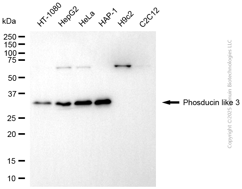
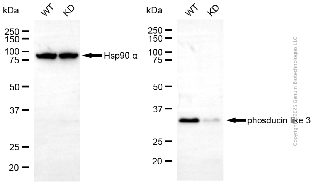
| Calculated MW | Predicted, 28 kDa, observed, 35 kDa |
| Gene Name | PDCL3 |
| Aliases | PDCL3; Phosducin Like 3; VIAF1; Viral IAP-Associated Factor 1; Phosducin-Like Protein 3; HTPHLP; VIAF-1; PhPL3; IAP-Associated Factor VIAF1; PHLP2A; PhLP2A; PHLP3; VIAF |
| Immunogen | Recombinant protein of human PDCL3 |
| Gene ID | 79031 |
|---|---|
| Other Names | Phosducin-like protein 3, HTPHLP, PhPL3, Viral IAP-associated factor 1, VIAF-1, PDCL3, PhLP2A, VIAF1 |
| Name | PDCL3 |
|---|---|
| Synonyms | PhLP2A, VIAF1 |
| Function | Acts as a chaperone for the angiogenic VEGF receptor KDR/VEGFR2, increasing its abundance by inhibiting its ubiquitination and degradation (PubMed:23792958, PubMed:26059764). Inhibits the folding activity of the chaperonin-containing T-complex (CCT) which leads to inhibition of cytoskeletal actin folding (PubMed:17429077). Acts as a chaperone during heat shock alongside HSP90 and HSP40/70 chaperone complexes (By similarity). Modulates the activation of caspases during apoptosis (PubMed:15371430). |
| Cellular Location | Cytoplasm. Cytoplasm, perinuclear region. Endoplasmic reticulum |
| Tissue Location | Expressed in endothelial cells (at protein level) (PubMed:26059764). Expressed in all tissues examined including spleen, thymus, prostate, testis, ovary, small intestine and colon (PubMed:15371430). |
Research Areas
Citations (0)

Thousands of laboratories across the world have published research that depended on the performance of antibodies from Abcepta to advance their research. Check out links to articles that cite our products in major peer-reviewed journals, organized by research category.
Submit your citation using an Abcepta antibody to
info@abcepta.com, and receive a free "I Love Antibodies" mug.
info@abcepta.com, and receive a free "I Love Antibodies" mug.

Application Protocols
Provided below are standard protocols that you may find useful for product applications.
Abcepta welcomes feedback from its customers.
If you have used an Abcepta product and would like to share how it has performed, please click on the "Submit Review" button and provide the requested information. Our staff will examine and post your review and contact you if needed.
If you have any additional inquiries please email technical services at tech@abcepta.com.
$ 399.20

$ 149.00
Cat# AGI2173
Ordering Information
United States
AlbaniaAustraliaAustriaBelgiumBosnia & HerzegovinaBrazilBulgariaCanadaCentral AmericaChinaCroatiaCyprusCzech RepublicDenmarkEstoniaFinlandFranceGermanyGreeceHong KongHungaryIcelandIndiaIndonesiaIrelandIsraelItalyJapanLatviaLithuaniaLuxembourgMacedoniaMalaysiaMaltaMexicoNetherlandsNew ZealandNorwayPakistanPolandPortugalRomaniaSerbiaSingaporeSlovakiaSloveniaSouth AfricaSouth KoreaSpainSwedenSwitzerlandTaiwanTurkeyUnited KingdomUnited StatesVietnamWorldwideOthers
USA Headquarters
(888) 735-7227 / (858) 622-0099 or (858) 875-1900
Other Products
Shipping Information
Domestic orders (in stock items)
Shipped out the same day. Orders placed after 1 PM (PST) will ship out the next business day.
International orders
Contact your local distributors
