


 Foundational characteristics of cancer include proliferation, angiogenesis, migration, evasion of apoptosis, and cellular immortality. Find key markers for these cellular processes and antibodies to detect them.
 The SUMOplot™ Analysis Program predicts and scores sumoylation sites in your protein. SUMOylation is a post-translational modification involved in various cellular processes, such as nuclear-cytosolic transport, transcriptional regulation, apoptosis, protein stability, response to stress, and progression through the cell cycle.
 The Autophagy Receptor Motif Plotter predicts and scores autophagy receptor binding sites in your protein. Identifying proteins connected to this pathway is critical to understanding the role of autophagy in physiological as well as pathological processes such as development, differentiation, neurodegenerative diseases, stress, infection, and cancer.
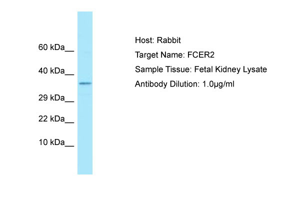
FCER2 Antibody - C-terminal region
Rabbit Polyclonal Antibody
- SPECIFICATION
- CITATIONS
- PROTOCOLS
- BACKGROUND

Application
| WB |
|---|---|
| Primary Accession | P06734 |
| Other Accession | NM_002002, NP_001993 |
| Reactivity | Human |
| Predicted | Human |
| Host | Rabbit |
| Clonality | Polyclonal |
| Calculated MW | 35kDa |
| Gene ID | 2208 |
|---|---|
| Alias Symbol | CD23, CD23A, CLEC4J, FCE2, IGEBF |
| Other Names | Low affinity immunoglobulin epsilon Fc receptor, BLAST-2, C-type lectin domain family 4 member J, Fc-epsilon-RII, Immunoglobulin E-binding factor, Lymphocyte IgE receptor, CD23, Low affinity immunoglobulin epsilon Fc receptor membrane-bound form, Low affinity immunoglobulin epsilon Fc receptor soluble form, FCER2, CD23A, CLEC4J, FCE2, IGEBF |
| Format | Liquid. Purified antibody supplied in 1x PBS buffer with 0.09% (w/v) sodium azide and 2% sucrose. |
| Reconstitution & Storage | Add 50 ul of distilled water. Final anti-FCER2 antibody concentration is 1 mg/ml in PBS buffer with 2% sucrose. For longer periods of storage, store at 20°C. Avoid repeat freeze-thaw cycles. |
| Precautions | FCER2 Antibody - C-terminal region is for research use only and not for use in diagnostic or therapeutic procedures. |
| Name | FCER2 |
|---|---|
| Synonyms | CD23A, CLEC4J, FCE2, IGEBF |
| Function | Low-affinity receptor for immunoglobulin E (IgE) and CR2/CD21. Has essential roles in the regulation of IgE production and in the differentiation of B cells. On B cells, initiates IgE-dependent antigen uptake and presentation to T cells (PubMed:2167225). On macrophages, upon IgE binding and antigen cross-linking induces intracellular killing of parasites through activation of L-Arginine- nitric oxide pathway (PubMed:7544003). |
| Cellular Location | Cell membrane; Single-pass type II membrane protein. Cell membrane; Lipid-anchor. Secreted. Note=Also exists as a soluble excreted form, sCD23 |
| Tissue Location | Detected in urine (at protein level). |

Thousands of laboratories across the world have published research that depended on the performance of antibodies from Abcepta to advance their research. Check out links to articles that cite our products in major peer-reviewed journals, organized by research category.
info@abcepta.com, and receive a free "I Love Antibodies" mug.

Provided below are standard protocols that you may find useful for product applications.
References
Ikuta K.,et al.Proc. Natl. Acad. Sci. U.S.A. 84:819-823(1987).
Kikutani H.,et al.Cell 47:657-665(1986).
Luedin C.,et al.EMBO J. 6:109-114(1987).
Grimwood J.,et al.Nature 428:529-535(2004).
Yokota A.,et al.Cell 55:611-618(1988).
If you have used an Abcepta product and would like to share how it has performed, please click on the "Submit Review" button and provide the requested information. Our staff will examine and post your review and contact you if needed.
If you have any additional inquiries please email technical services at tech@abcepta.com.
Ordering Information
Other Products
Shipping Information
