


 Foundational characteristics of cancer include proliferation, angiogenesis, migration, evasion of apoptosis, and cellular immortality. Find key markers for these cellular processes and antibodies to detect them.
 The SUMOplot™ Analysis Program predicts and scores sumoylation sites in your protein. SUMOylation is a post-translational modification involved in various cellular processes, such as nuclear-cytosolic transport, transcriptional regulation, apoptosis, protein stability, response to stress, and progression through the cell cycle.
 The Autophagy Receptor Motif Plotter predicts and scores autophagy receptor binding sites in your protein. Identifying proteins connected to this pathway is critical to understanding the role of autophagy in physiological as well as pathological processes such as development, differentiation, neurodegenerative diseases, stress, infection, and cancer.
GPR34 Antibody - C-terminal region
Rabbit Polyclonal Antibody
- SPECIFICATION
- CITATIONS
- PROTOCOLS
- BACKGROUND

Application
| WB |
|---|---|
| Primary Accession | Q9UPC5 |
| Other Accession | NM_005300, NP_005291 |
| Reactivity | Human, Mouse, Rat, Rabbit, Horse, Bovine, Guinea Pig, Dog |
| Predicted | Human, Mouse, Rat, Rabbit, Pig, Horse, Bovine, Guinea Pig, Dog |
| Host | Rabbit |
| Clonality | Polyclonal |
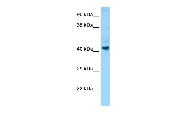
| Calculated MW | 44kDa |
| Gene ID | 2857 |
|---|---|
| Other Names | Probable G-protein coupled receptor 34, GPR34 |
| Format | Liquid. Purified antibody supplied in 1x PBS buffer with 0.09% (w/v) sodium azide and 2% sucrose. |
| Reconstitution & Storage | Add 50 ul of distilled water. Final anti-GPR34 antibody concentration is 1 mg/ml in PBS buffer with 2% sucrose. For longer periods of storage, store at 20°C. Avoid repeat freeze-thaw cycles. |
| Precautions | GPR34 Antibody - C-terminal region is for research use only and not for use in diagnostic or therapeutic procedures. |
| Name | GPR34 |
|---|---|
| Function | G-protein-coupled receptor of lysophosphatidylserine (LysoPS) that plays different roles in immune response (PubMed:16460680). Acts a damage-sensing receptor that triggers tissue repair upon recognition of dying neutrophils (By similarity). Mechanistically, apoptotic neutrophils release lysophosphatydilserine that are recognized by type 3 innate lymphoid cells (ILC3s) via GPR34, which activates downstream PI3K-AKT and RAS-ERK signaling pathways leading to STAT3 activation and IL-22 production (By similarity). Plays an important role in microglial function, controlling morphology and phagocytosis (By similarity). |
| Cellular Location | Cell membrane; Multi-pass membrane protein |
| Tissue Location | Broadly expressed. Highly expressed on mast cells (PubMed:16460680). |

Thousands of laboratories across the world have published research that depended on the performance of antibodies from Abcepta to advance their research. Check out links to articles that cite our products in major peer-reviewed journals, organized by research category.
info@abcepta.com, and receive a free "I Love Antibodies" mug.

Provided below are standard protocols that you may find useful for product applications.
References
Schoneberg T.,et al.Biochim. Biophys. Acta 1446:57-70(1999).
Marchese A.,et al.Genomics 56:12-21(1999).
Jacobi F.K.,et al.Hum. Genet. 107:89-91(2000).
Ota T.,et al.Nat. Genet. 36:40-45(2004).
If you have used an Abcepta product and would like to share how it has performed, please click on the "Submit Review" button and provide the requested information. Our staff will examine and post your review and contact you if needed.
If you have any additional inquiries please email technical services at tech@abcepta.com.
Ordering Information
Other Products
Shipping Information
