


 Foundational characteristics of cancer include proliferation, angiogenesis, migration, evasion of apoptosis, and cellular immortality. Find key markers for these cellular processes and antibodies to detect them.
 The SUMOplot™ Analysis Program predicts and scores sumoylation sites in your protein. SUMOylation is a post-translational modification involved in various cellular processes, such as nuclear-cytosolic transport, transcriptional regulation, apoptosis, protein stability, response to stress, and progression through the cell cycle.
 The Autophagy Receptor Motif Plotter predicts and scores autophagy receptor binding sites in your protein. Identifying proteins connected to this pathway is critical to understanding the role of autophagy in physiological as well as pathological processes such as development, differentiation, neurodegenerative diseases, stress, infection, and cancer.
NOS1 Antibody
Purified Mouse Monoclonal Antibody
- SPECIFICATION
- CITATIONS
- PROTOCOLS
- BACKGROUND

Application
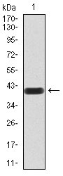
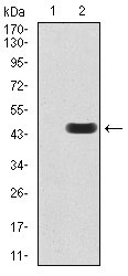
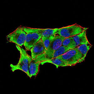
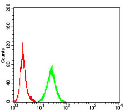
| WB, FC, ICC, E |
|---|---|
| Primary Accession | P29475 |
| Reactivity | Human |
| Host | Mouse |
| Clonality | Monoclonal |
| Clone Names | 2E11G6 |
| Isotype | IgG1 |
| Calculated MW | 161kDa |
| Description | The protein encoded by this gene belongs to the family of nitric oxide synthases, which synthesize nitric oxide from L-arginine. Nitric oxide is a reactive free radical, which acts as a biologic mediator in several processes, including neurotransmission, and antimicrobial and antitumoral activities. In the brain and peripheral nervous system, nitric oxide displays many properties of a neurotransmitter, and has been implicated in neurotoxicity associated with stroke and neurodegenerative diseases, neural regulation of smooth muscle, including peristalsis, and penile erection. This protein is ubiquitously expressed, with high level of expression in skeletal muscle. Multiple transcript variants that differ in the 5' UTR have been described for this gene but the full-length nature of these transcripts is not known. Additionally, alternatively spliced transcript variants encoding different isoforms (some testis-specific) have been found for this gene. |
| Immunogen | Purified recombinant fragment of human NOS1 (AA: 17-153) expressed in E. Coli. |
| Formulation | Purified antibody in PBS with 0.05% sodium azide |
| Gene ID | 4842 |
|---|---|
| Other Names | Nitric oxide synthase, brain, 1.14.13.39, Constitutive NOS, NC-NOS, NOS type I, Neuronal NOS, N-NOS, nNOS, Peptidyl-cysteine S-nitrosylase NOS1, bNOS, NOS1 |
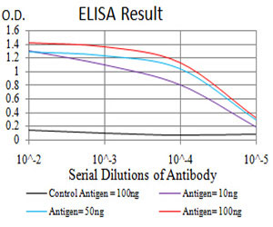
| Dilution | WB~~1/500 - 1/2000 FC~~1/200 - 1/400 ICC~~N/A E~~1/10000 |
| Storage | Maintain refrigerated at 2-8°C for up to 6 months. For long term storage store at -20°C in small aliquots to prevent freeze-thaw cycles. |
| Precautions | NOS1 Antibody is for research use only and not for use in diagnostic or therapeutic procedures. |
| Name | NOS1 (HGNC:7872) |
|---|---|
| Function | Produces nitric oxide (NO) which is a messenger molecule with diverse functions throughout the body. In the brain and peripheral nervous system, NO displays many properties of a neurotransmitter. Probably has nitrosylase activity and mediates cysteine S-nitrosylation of cytoplasmic target proteins such SRR. |
| Cellular Location | Cell membrane, sarcolemma {ECO:0000250|UniProtKB:Q9Z0J4}; Peripheral membrane protein. Cell projection, dendritic spine {ECO:0000250|UniProtKB:P29476}. Note=In skeletal muscle, it is localized beneath the sarcolemma of fast-twitch muscle fiber by associating with the dystrophin glycoprotein complex (By similarity) In neurons, enriched in dendritic spines (By similarity) {ECO:0000250|UniProtKB:P29476, ECO:0000250|UniProtKB:Q9Z0J4} |
| Tissue Location | Isoform 1 is ubiquitously expressed: detected in skeletal muscle and brain, also in testis, lung and kidney, and at low levels in heart, adrenal gland and retina. Not detected in the platelets. Isoform 3 is expressed only in testis. Isoform 4 is detected in testis, skeletal muscle, lung, and kidney, at low levels in the brain, but not in the heart and adrenal gland |

Thousands of laboratories across the world have published research that depended on the performance of antibodies from Abcepta to advance their research. Check out links to articles that cite our products in major peer-reviewed journals, organized by research category.
info@abcepta.com, and receive a free "I Love Antibodies" mug.

Provided below are standard protocols that you may find useful for product applications.
References
1.J Neurochem. 2013 Aug;126(3):318-30.2.J Leukoc Biol. 2012 Jun;91(6):947-56.
If you have used an Abcepta product and would like to share how it has performed, please click on the "Submit Review" button and provide the requested information. Our staff will examine and post your review and contact you if needed.
If you have any additional inquiries please email technical services at tech@abcepta.com.
Ordering Information
Other Products
Shipping Information
