


 Foundational characteristics of cancer include proliferation, angiogenesis, migration, evasion of apoptosis, and cellular immortality. Find key markers for these cellular processes and antibodies to detect them.
 The SUMOplot™ Analysis Program predicts and scores sumoylation sites in your protein. SUMOylation is a post-translational modification involved in various cellular processes, such as nuclear-cytosolic transport, transcriptional regulation, apoptosis, protein stability, response to stress, and progression through the cell cycle.
 The Autophagy Receptor Motif Plotter predicts and scores autophagy receptor binding sites in your protein. Identifying proteins connected to this pathway is critical to understanding the role of autophagy in physiological as well as pathological processes such as development, differentiation, neurodegenerative diseases, stress, infection, and cancer.
MAN2A1 Antibody (Center)
Affinity Purified Rabbit Polyclonal Antibody (Pab)
- SPECIFICATION
- CITATIONS
- PROTOCOLS
- BACKGROUND

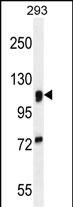
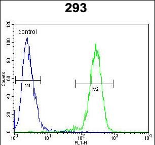
Application
| WB, FC, E |
|---|---|
| Primary Accession | Q16706 |
| Other Accession | NP_002363.2 |
| Reactivity | Human |
| Host | Rabbit |
| Clonality | Polyclonal |
| Isotype | Rabbit IgG |
| Calculated MW | 131141 Da |
| Antigen Region | 456-482 aa |
| Gene ID | 4124 |
|---|---|
| Other Names | Alpha-mannosidase 2, Golgi alpha-mannosidase II, AMan II, Man II, Mannosidase alpha class 2A member 1, Mannosyl-oligosaccharide 1, 3-1, 6-alpha-mannosidase, MAN2A1, MANA2 |
| Target/Specificity | This MAN2A1 antibody is generated from rabbits immunized with a KLH conjugated synthetic peptide between 456-482 amino acids from the Central region of human MAN2A1. |
| Dilution | WB~~1:1000 FC~~1:10~50 E~~Use at an assay dependent concentration. |
| Format | Purified polyclonal antibody supplied in PBS with 0.09% (W/V) sodium azide. This antibody is purified through a protein A column, followed by peptide affinity purification. |
| Storage | Maintain refrigerated at 2-8°C for up to 2 weeks. For long term storage store at -20°C in small aliquots to prevent freeze-thaw cycles. |
| Precautions | MAN2A1 Antibody (Center) is for research use only and not for use in diagnostic or therapeutic procedures. |
| Name | MAN2A1 |
|---|---|
| Synonyms | MANA2 |
| Function | Catalyzes the first committed step in the biosynthesis of complex N-glycans. It controls conversion of high mannose to complex N- glycans; the final hydrolytic step in the N-glycan maturation pathway. |
| Cellular Location | Golgi apparatus membrane {ECO:0000250|UniProtKB:P28494}; Single-pass type II membrane protein {ECO:0000250|UniProtKB:P28494} |

Thousands of laboratories across the world have published research that depended on the performance of antibodies from Abcepta to advance their research. Check out links to articles that cite our products in major peer-reviewed journals, organized by research category.
info@abcepta.com, and receive a free "I Love Antibodies" mug.

Provided below are standard protocols that you may find useful for product applications.
Background
This gene encodes a protein which is a member of family 38 of the glycosyl hydrolases. The protein is located in the Golgi and catalyzes the final hydrolytic step in the asparagine-linked oligosaccharide (N-glycan) maturation pathway. Mutations in the mouse homolog of this gene have been shown to cause a systemic autoimmune disease similar to human systemic lupus erythematosus.
References
Han, S., et al. Leuk. Res. 34(10):1271-1274(2010)
Rose, J.E., et al. Mol. Med. 16 (7-8), 247-253 (2010) :
Zhou, J.B., et al. Med. Sci. Monit. 16 (6), BR179-BR183 (2010) :
Miyagawa, H., et al. Rheumatology (Oxford) 47(2):158-164(2008)
Yang, Q., et al. BMC Med. Genet. 8 SUPPL 1, S12 (2007) :
If you have used an Abcepta product and would like to share how it has performed, please click on the "Submit Review" button and provide the requested information. Our staff will examine and post your review and contact you if needed.
If you have any additional inquiries please email technical services at tech@abcepta.com.
Ordering Information
Other Products
Shipping Information
