


 Foundational characteristics of cancer include proliferation, angiogenesis, migration, evasion of apoptosis, and cellular immortality. Find key markers for these cellular processes and antibodies to detect them.
 The SUMOplot™ Analysis Program predicts and scores sumoylation sites in your protein. SUMOylation is a post-translational modification involved in various cellular processes, such as nuclear-cytosolic transport, transcriptional regulation, apoptosis, protein stability, response to stress, and progression through the cell cycle.
 The Autophagy Receptor Motif Plotter predicts and scores autophagy receptor binding sites in your protein. Identifying proteins connected to this pathway is critical to understanding the role of autophagy in physiological as well as pathological processes such as development, differentiation, neurodegenerative diseases, stress, infection, and cancer.
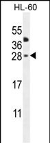
CLEC2A Antibody (Center)
Affinity Purified Rabbit Polyclonal Antibody (Pab)
- SPECIFICATION
- CITATIONS
- PROTOCOLS
- BACKGROUND

Application
| WB, E |
|---|---|
| Primary Accession | Q6UVW9 |
| Other Accession | NP_001124183.1 |
| Reactivity | Human |
| Host | Rabbit |
| Clonality | Polyclonal |
| Isotype | Rabbit IgG |
| Calculated MW | 19972 Da |
| Antigen Region | 79-107 aa |
| Gene ID | 387836 |
|---|---|
| Other Names | C-type lectin domain family 2 member A, Keratinocyte-associated C-type lectin, KACL, Proliferation-induced lymphocyte-associated receptor, PILAR, CLEC2A, KACL |
| Target/Specificity | This CLEC2A antibody is generated from rabbits immunized with a KLH conjugated synthetic peptide between 79-107 amino acids from the Central region of human CLEC2A. |
| Dilution | WB~~1:1000 E~~Use at an assay dependent concentration. |
| Format | Purified polyclonal antibody supplied in PBS with 0.09% (W/V) sodium azide. This antibody is purified through a protein A column, followed by peptide affinity purification. |
| Storage | Maintain refrigerated at 2-8°C for up to 2 weeks. For long term storage store at -20°C in small aliquots to prevent freeze-thaw cycles. |
| Precautions | CLEC2A Antibody (Center) is for research use only and not for use in diagnostic or therapeutic procedures. |
| Name | CLEC2A |
|---|---|
| Synonyms | KACL |
| Function | Membrane-bound protein expressed mainly on keratinocytes which acts as a ligand to stimulate the activating receptor NKp65/KLRF2, expressed on the surface of natural killer (NK) cells (PubMed:25510854). Facilitates thereby dedicated immune recognition of keratinocytes leading to natural killer cell mediated cytotoxicity (PubMed:20194751). Also plays a role in modulating the extent of T-cell expansion (PubMed:18550855). |
| Cellular Location | Cell membrane; Single-pass type II membrane protein |
| Tissue Location | Mainly expressed in skin. Also expressed in keratinocytes, spleen, thymus, small intestine, peripheral blood monocytes, bone marrow, ovary, testis and skin. High expression in CD8(+), B-lymphocytes and naive CD4(+) T-cells. Restricted mostly to proliferating lymphocytes. Not detected in myeloid leukocytes or natural killer (NK) cells. |

Thousands of laboratories across the world have published research that depended on the performance of antibodies from Abcepta to advance their research. Check out links to articles that cite our products in major peer-reviewed journals, organized by research category.
info@abcepta.com, and receive a free "I Love Antibodies" mug.

Provided below are standard protocols that you may find useful for product applications.
Background
CLEC2A belongs to the CLEC2 family of activation-induced, natural killer gene complex-encoded C-type lectin-like receptors (Spreu et al., 2007 [PubMed 18046548]).
References
Spreu, J., et al. Proc. Natl. Acad. Sci. U.S.A. 107(11):5100-5105(2010)
Ozaki, Y., et al. J. Thromb. Haemost. 7 SUPPL 1, 191-194 (2009) :
Huarte, E., et al. Blood 112(4):1259-1268(2008)
Spreu, J., et al. Immunogenetics 59(12):903-912(2007)
Clark, H.F., et al. Genome Res. 13(10):2265-2270(2003)
If you have used an Abcepta product and would like to share how it has performed, please click on the "Submit Review" button and provide the requested information. Our staff will examine and post your review and contact you if needed.
If you have any additional inquiries please email technical services at tech@abcepta.com.
Ordering Information
Other Products
Shipping Information
