


 Foundational characteristics of cancer include proliferation, angiogenesis, migration, evasion of apoptosis, and cellular immortality. Find key markers for these cellular processes and antibodies to detect them.
 The SUMOplot™ Analysis Program predicts and scores sumoylation sites in your protein. SUMOylation is a post-translational modification involved in various cellular processes, such as nuclear-cytosolic transport, transcriptional regulation, apoptosis, protein stability, response to stress, and progression through the cell cycle.
 The Autophagy Receptor Motif Plotter predicts and scores autophagy receptor binding sites in your protein. Identifying proteins connected to this pathway is critical to understanding the role of autophagy in physiological as well as pathological processes such as development, differentiation, neurodegenerative diseases, stress, infection, and cancer.
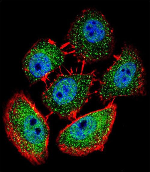
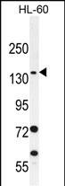
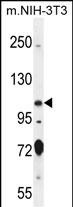
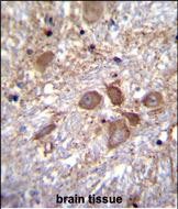
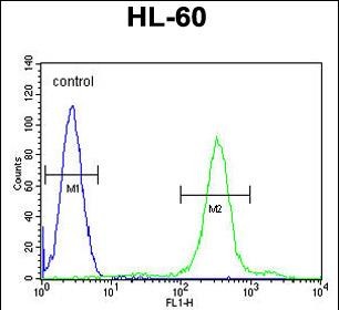
SPECC1 Antibody (Center)
Affinity Purified Rabbit Polyclonal Antibody (Pab)
- SPECIFICATION
- CITATIONS
- PROTOCOLS
- BACKGROUND

Application
| WB, IF, FC, IHC-P, E |
|---|---|
| Primary Accession | Q69YQ0 |
| Other Accession | Q2KN98, Q2KN93, Q2KN97, NP_001138940.1 |
| Reactivity | Human, Mouse |
| Predicted | Chicken, Zebrafish |
| Host | Rabbit |
| Clonality | Polyclonal |
| Isotype | Rabbit IgG |
| Calculated MW | 124544 Da |
| Antigen Region | 705-733 aa |
| Gene ID | 23384 |
|---|---|
| Other Names | Cytospin-A, Renal carcinoma antigen NY-REN-22, Sperm antigen with calponin homology and coiled-coil domains 1-like, SPECC1-like protein, SPECC1L, CYTSA, KIAA0376 |
| Target/Specificity | This SPECC1 antibody is generated from rabbits immunized with a KLH conjugated synthetic peptide between 705-733 amino acids from the Central region of human SPECC1. |
| Dilution | WB~~1:1000 IF~~1:10~50 FC~~1:10~50 IHC-P~~1:10~50 E~~Use at an assay dependent concentration. |
| Format | Purified polyclonal antibody supplied in PBS with 0.09% (W/V) sodium azide. This antibody is purified through a protein A column, followed by peptide affinity purification. |
| Storage | Maintain refrigerated at 2-8°C for up to 2 weeks. For long term storage store at -20°C in small aliquots to prevent freeze-thaw cycles. |
| Precautions | SPECC1 Antibody (Center) is for research use only and not for use in diagnostic or therapeutic procedures. |
| Name | SPECC1L |
|---|---|
| Synonyms | CYTSA, KIAA0376 |
| Function | Involved in cytokinesis and spindle organization. May play a role in actin cytoskeleton organization and microtubule stabilization and hence required for proper cell adhesion and migration. |
| Cellular Location | Cytoplasm, cytoskeleton. Cytoplasm, cytoskeleton, spindle. Cell junction, gap junction. Note=Colocalizes with acetylated alpha- tubulin, gamma-tubulin and F-actin. Also observed in a ring around gamma-tubulin containing centrioles possibly in the microtubule organizing center |

Thousands of laboratories across the world have published research that depended on the performance of antibodies from Abcepta to advance their research. Check out links to articles that cite our products in major peer-reviewed journals, organized by research category.
info@abcepta.com, and receive a free "I Love Antibodies" mug.

Provided below are standard protocols that you may find useful for product applications.
Background
CYTSAis involved in cytokinesis and spindle organization.
References
Scanlan, M.J., et al. Int. J. Cancer 83(4):456-464(1999)
If you have used an Abcepta product and would like to share how it has performed, please click on the "Submit Review" button and provide the requested information. Our staff will examine and post your review and contact you if needed.
If you have any additional inquiries please email technical services at tech@abcepta.com.
Ordering Information
Other Products
Shipping Information
