


 Foundational characteristics of cancer include proliferation, angiogenesis, migration, evasion of apoptosis, and cellular immortality. Find key markers for these cellular processes and antibodies to detect them.
 The SUMOplot™ Analysis Program predicts and scores sumoylation sites in your protein. SUMOylation is a post-translational modification involved in various cellular processes, such as nuclear-cytosolic transport, transcriptional regulation, apoptosis, protein stability, response to stress, and progression through the cell cycle.
 The Autophagy Receptor Motif Plotter predicts and scores autophagy receptor binding sites in your protein. Identifying proteins connected to this pathway is critical to understanding the role of autophagy in physiological as well as pathological processes such as development, differentiation, neurodegenerative diseases, stress, infection, and cancer.
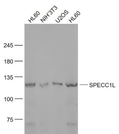
SPECC1L Polyclonal Antibody
Purified Rabbit Polyclonal Antibody (Pab)
- SPECIFICATION
- CITATIONS
- PROTOCOLS
- BACKGROUND

Application
| WB, IHC-P, IHC-F, IF, E |
|---|---|
| Primary Accession | Q69YQ0 |
| Reactivity | Rat, Pig, Dog, Bovine |
| Host | Rabbit |
| Clonality | Polyclonal |
| Calculated MW | 125 KDa |
| Physical State | Liquid |
| Immunogen | KLH conjugated synthetic peptide derived from human SPECC1L |
| Epitope Specificity | 161-260/1117 |
| Isotype | IgG |
| Purity | affinity purified by Protein A |
| Buffer | 0.01M TBS (pH7.4) with 1% BSA, 0.02% Proclin300 and 50% Glycerol. |
| SUBCELLULAR LOCATION | Cytoplasm, cytoskeleton. Cytoplasm, cytoskeleton, spindle. Cell junction, gap junction. Note=Colocalizes with acetylated alpha-tubulin, gamma-tubulin and F-actin. Also observed in a ring around gamma-tubulin containing centrioles possibly in the microtubule organizing center. |
| SIMILARITY | Belongs to the cytospin-A family. Contains 1 CH (calponin-homology) domain. |
| SUBUNIT | May interact with both microtubules and actin cytoskeleton. |
| DISEASE | Defects in SPECC1L are the cause of facial clefting oblique type 1 (OBLFC1) [MIM:600251]. A rare form of facial clefting. A facial cleft is any of the fissures between the embryonic prominences that normally unite to form the face. |
| Important Note | This product as supplied is intended for research use only, not for use in human, therapeutic or diagnostic applications. |
| Background Descriptions | This gene encodes a coiled-coil domain containing protein. The encoded protein may play a critical role in actin-cytoskeletal reorganization during facial morphogenesis. Mutations in this gene are a cause of oblique facial clefting-1. Alternatively spliced transcript variants encoding multiple isoforms have been observed for this gene. A read-through transcript composed of SPECC1L (sperm antigen with calponin homology and coiled-coil domains 1-like) and the downstream ADORA2A (adenosine A2a receptor) gene sequence has been identified, but it is thought to be non-coding. [provided by RefSeq, Jun 2013] |
| Gene ID | 23384 |
|---|---|
| Other Names | Cytospin-A, Renal carcinoma antigen NY-REN-22, Sperm antigen with calponin homology and coiled-coil domains 1-like, SPECC1-like protein, SPECC1L, CYTSA, KIAA0376 |
| Dilution | WB=1:500-2000,IHC-P=1:100-500,IHC-F=1:100-500,IF=1:100-500,ELISA=1:5000-10000 |
| Storage | Store at -20 ℃ for one year. Avoid repeated freeze/thaw cycles. When reconstituted in sterile pH 7.4 0.01M PBS or diluent of antibody the antibody is stable for at least two weeks at 2-4 ℃. |
| Name | SPECC1L |
|---|---|
| Synonyms | CYTSA, KIAA0376 |
| Function | Involved in cytokinesis and spindle organization. May play a role in actin cytoskeleton organization and microtubule stabilization and hence required for proper cell adhesion and migration. |
| Cellular Location | Cytoplasm, cytoskeleton. Cytoplasm, cytoskeleton, spindle. Cell junction, gap junction. Note=Colocalizes with acetylated alpha- tubulin, gamma-tubulin and F-actin. Also observed in a ring around gamma-tubulin containing centrioles possibly in the microtubule organizing center |
Research Areas
Citations (0)

Thousands of laboratories across the world have published research that depended on the performance of antibodies from Abcepta to advance their research. Check out links to articles that cite our products in major peer-reviewed journals, organized by research category.
Submit your citation using an Abcepta antibody to
info@abcepta.com, and receive a free "I Love Antibodies" mug.
info@abcepta.com, and receive a free "I Love Antibodies" mug.

Application Protocols
Provided below are standard protocols that you may find useful for product applications.
Abcepta welcomes feedback from its customers.
If you have used an Abcepta product and would like to share how it has performed, please click on the "Submit Review" button and provide the requested information. Our staff will examine and post your review and contact you if needed.
If you have any additional inquiries please email technical services at tech@abcepta.com.
$ 385.00
Cat# AP58749
Ordering Information
United States
AlbaniaAustraliaAustriaBelgiumBosnia & HerzegovinaBrazilBulgariaCanadaCentral AmericaChinaCroatiaCyprusCzech RepublicDenmarkEstoniaFinlandFranceGermanyGreeceHong KongHungaryIcelandIndiaIndonesiaIrelandIsraelItalyJapanLatviaLithuaniaLuxembourgMacedoniaMalaysiaMaltaMexicoNetherlandsNew ZealandNorwayPakistanPolandPortugalRomaniaSerbiaSingaporeSlovakiaSloveniaSouth AfricaSouth KoreaSpainSwedenSwitzerlandTaiwanTurkeyUnited KingdomUnited StatesVietnamWorldwideOthers
USA Headquarters
(888) 735-7227 / (858) 622-0099 or (858) 875-1900
Other Products
Shipping Information
Domestic orders (in stock items)
Shipped out the same day. Orders placed after 1 PM (PST) will ship out the next business day.
International orders
Contact your local distributors
