


 Foundational characteristics of cancer include proliferation, angiogenesis, migration, evasion of apoptosis, and cellular immortality. Find key markers for these cellular processes and antibodies to detect them.
 The SUMOplot™ Analysis Program predicts and scores sumoylation sites in your protein. SUMOylation is a post-translational modification involved in various cellular processes, such as nuclear-cytosolic transport, transcriptional regulation, apoptosis, protein stability, response to stress, and progression through the cell cycle.
 The Autophagy Receptor Motif Plotter predicts and scores autophagy receptor binding sites in your protein. Identifying proteins connected to this pathway is critical to understanding the role of autophagy in physiological as well as pathological processes such as development, differentiation, neurodegenerative diseases, stress, infection, and cancer.
NRBP1 Antibody (N-term)
Affinity Purified Rabbit Polyclonal Antibody (Pab)
- SPECIFICATION
- CITATIONS
- PROTOCOLS
- BACKGROUND

Application
| WB, E |
|---|---|
| Primary Accession | Q9UHY1 |
| Other Accession | Q99J45, Q4R8X0, NP_037524.1 |
| Reactivity | Human |
| Predicted | Monkey, Mouse |
| Host | Rabbit |
| Clonality | Polyclonal |
| Isotype | Rabbit IgG |
| Calculated MW | 59845 Da |
| Antigen Region | 86-115 aa |
| Gene ID | 29959 |
|---|---|
| Other Names | Nuclear receptor-binding protein, NRBP1, BCON3 {ECO:0000312|EMBL:AAF219671}, NRBP |
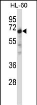
| Target/Specificity | This NRBP1 antibody is generated from rabbits immunized with a KLH conjugated synthetic peptide between 86-115 amino acids from the N-terminal region of human NRBP1. |
| Dilution | WB~~1:1000 E~~Use at an assay dependent concentration. |
| Format | Purified polyclonal antibody supplied in PBS with 0.09% (W/V) sodium azide. This antibody is purified through a protein A column, followed by peptide affinity purification. |
| Storage | Maintain refrigerated at 2-8°C for up to 2 weeks. For long term storage store at -20°C in small aliquots to prevent freeze-thaw cycles. |
| Precautions | NRBP1 Antibody (N-term) is for research use only and not for use in diagnostic or therapeutic procedures. |
| Name | NRBP1 |
|---|---|
| Synonyms | BCON3 {ECO:0000312|EMBL:AAF21967.1}, NRB |
| Function | Required for embryonic development (By similarity). Plays a role in intestinal epithelial cell fate and proliferation, thereby involved in the architectural development of the intestine potentially via the regulation of Wnt-responsive genes (By similarity). May play a role in subcellular trafficking between the endoplasmic reticulum and Golgi apparatus through interactions with the Rho-type GTPases (PubMed:11956649). Binding to the NS3 protein of dengue virus type 2 appears to subvert this activity into the alteration of the intracellular membrane structure associated with flaviviral replication (PubMed:15084397). |
| Cellular Location | Cytoplasm, cell cortex. Endomembrane system. Cell projection, lamellipodium. Note=Colocalizes with activated RAC3 to endomembranes and at the cell periphery in lamellipodia |
| Tissue Location | Ubiquitously expressed in all tissues examined with high levels in the testis. |

Thousands of laboratories across the world have published research that depended on the performance of antibodies from Abcepta to advance their research. Check out links to articles that cite our products in major peer-reviewed journals, organized by research category.
info@abcepta.com, and receive a free "I Love Antibodies" mug.

Provided below are standard protocols that you may find useful for product applications.
Background
NRBP1 may play a role in subcellular trafficking between the endoplasmic reticulum and Golgi apparatus through interactions with the Rho-type GTPases. Binding to the NS3 protein of dengue virus type 2 appears to subvert this activity into the alteration of the intracellular membrane structure associated with flaviviral replication.
References
Ewing, R.M., et al. Mol. Syst. Biol. 3, 89 (2007) :
Olsen, J.V., et al. Cell 127(3):635-648(2006)
Wang, H., et al. FEBS Lett. 580(25):6015-6021(2006)
Beausoleil, S.A., et al. Nat. Biotechnol. 24(10):1285-1292(2006)
Hillier, L.W., et al. Nature 434(7034):724-731(2005)
If you have used an Abcepta product and would like to share how it has performed, please click on the "Submit Review" button and provide the requested information. Our staff will examine and post your review and contact you if needed.
If you have any additional inquiries please email technical services at tech@abcepta.com.
Ordering Information
Other Products
Shipping Information
