


 Foundational characteristics of cancer include proliferation, angiogenesis, migration, evasion of apoptosis, and cellular immortality. Find key markers for these cellular processes and antibodies to detect them.
 The SUMOplot™ Analysis Program predicts and scores sumoylation sites in your protein. SUMOylation is a post-translational modification involved in various cellular processes, such as nuclear-cytosolic transport, transcriptional regulation, apoptosis, protein stability, response to stress, and progression through the cell cycle.
 The Autophagy Receptor Motif Plotter predicts and scores autophagy receptor binding sites in your protein. Identifying proteins connected to this pathway is critical to understanding the role of autophagy in physiological as well as pathological processes such as development, differentiation, neurodegenerative diseases, stress, infection, and cancer.
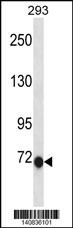
CLIC6 Antibody(C-term)
Affinity Purified Rabbit Polyclonal Antibody (Pab)
- SPECIFICATION
- CITATIONS
- PROTOCOLS
- BACKGROUND

Application
| WB, E |
|---|---|
| Primary Accession | Q96NY7 |
| Other Accession | Q9N2G5, NP_444507.1 |
| Reactivity | Human |
| Predicted | Rabbit |
| Host | Rabbit |
| Clonality | Polyclonal |
| Isotype | Rabbit IgG |
| Calculated MW | 73012 Da |
| Antigen Region | 676-704 aa |
| Gene ID | 54102 |
|---|---|
| Other Names | Chloride intracellular channel protein 6, Parchorin, CLIC6, CLIC1L |
| Target/Specificity | This CLIC6 antibody is generated from rabbits immunized with a KLH conjugated synthetic peptide between 676-704 amino acids from the C-terminal region of human CLIC6. |
| Dilution | WB~~1:1000 E~~Use at an assay dependent concentration. |
| Format | Purified polyclonal antibody supplied in PBS with 0.09% (W/V) sodium azide. This antibody is purified through a protein A column, followed by peptide affinity purification. |
| Storage | Maintain refrigerated at 2-8°C for up to 2 weeks. For long term storage store at -20°C in small aliquots to prevent freeze-thaw cycles. |
| Precautions | CLIC6 Antibody(C-term) is for research use only and not for use in diagnostic or therapeutic procedures. |
| Name | CLIC6 {ECO:0000303|PubMed:37838179, ECO:0000312|HGNC:HGNC:2065} |
|---|---|
| Function | In the soluble state, catalyzes glutaredoxin-like thiol disulfide exchange reactions with reduced glutathione as electron donor (By similarity). Can insert into membranes and form voltage-dependent chloride-selective channels. The channel opens upon membrane depolarization at positive voltages and closes at negative membrane voltages (PubMed:37838179). May play a critical role in water-secreting cells, possibly through the regulation of chloride ion transport (By similarity). |
| Cellular Location | Cytoplasm. Cell membrane; Single-pass membrane protein Note=Predominantly cytoplasmic. Upon chloride ion efflux from the cell, it is translocated to the plasma membrane (By similarity) |
| Tissue Location | Expressed in brain, placenta, pancreas, liver, lung, heart, kidney, liver, spleen, soleus muscle, and brown fat |

Thousands of laboratories across the world have published research that depended on the performance of antibodies from Abcepta to advance their research. Check out links to articles that cite our products in major peer-reviewed journals, organized by research category.
info@abcepta.com, and receive a free "I Love Antibodies" mug.

Provided below are standard protocols that you may find useful for product applications.
Background
This gene encodes a member of the chloride intracellular channel family of proteins. The gene is part of a large triplicated region found on chromosomes 1, 6, and 21. An alternatively spliced transcript variant has been described, but its biological validity has not been determined.
References
Wheeler, H.E., et al. PLoS Genet. 5 (10), E1000685 (2009) :
Friedli, M., et al. Gene 320, 31-40 (2003) :
Strippoli, P., et al. Mamm. Genome 13(8):456-462(2002)
Scanlan, M.J., et al. Cancer Immun. 1, 4 (2001) :
If you have used an Abcepta product and would like to share how it has performed, please click on the "Submit Review" button and provide the requested information. Our staff will examine and post your review and contact you if needed.
If you have any additional inquiries please email technical services at tech@abcepta.com.
Ordering Information
Other Products
Shipping Information
