


 Foundational characteristics of cancer include proliferation, angiogenesis, migration, evasion of apoptosis, and cellular immortality. Find key markers for these cellular processes and antibodies to detect them.
 The SUMOplot™ Analysis Program predicts and scores sumoylation sites in your protein. SUMOylation is a post-translational modification involved in various cellular processes, such as nuclear-cytosolic transport, transcriptional regulation, apoptosis, protein stability, response to stress, and progression through the cell cycle.
 The Autophagy Receptor Motif Plotter predicts and scores autophagy receptor binding sites in your protein. Identifying proteins connected to this pathway is critical to understanding the role of autophagy in physiological as well as pathological processes such as development, differentiation, neurodegenerative diseases, stress, infection, and cancer.
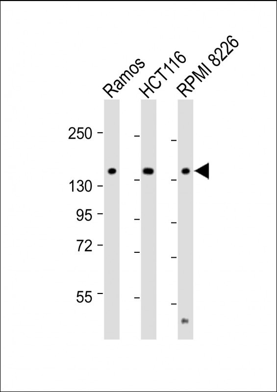
TIAM1 Antibody (C-term)
Affinity Purified Rabbit Polyclonal Antibody (Pab)
- SPECIFICATION
- CITATIONS
- PROTOCOLS
- BACKGROUND

Application
| WB, E |
|---|---|
| Primary Accession | Q13009 |
| Other Accession | Q60610, NP_003244.2 |
| Reactivity | Human |
| Predicted | Mouse |
| Host | Rabbit |
| Clonality | Polyclonal |
| Isotype | Rabbit IgG |
| Calculated MW | 177508 Da |
| Antigen Region | 1478-1506 aa |
| Gene ID | 7074 |
|---|---|
| Other Names | T-lymphoma invasion and metastasis-inducing protein 1, TIAM-1, TIAM1 |
| Target/Specificity | This TIAM1 antibody is generated from rabbits immunized with a KLH conjugated synthetic peptide between 1478-1506 amino acids from the C-terminal region of human TIAM1. |
| Dilution | WB~~1:2000 E~~Use at an assay dependent concentration. |
| Format | Purified polyclonal antibody supplied in PBS with 0.09% (W/V) sodium azide. This antibody is purified through a protein A column, followed by peptide affinity purification. |
| Storage | Maintain refrigerated at 2-8°C for up to 2 weeks. For long term storage store at -20°C in small aliquots to prevent freeze-thaw cycles. |
| Precautions | TIAM1 Antibody (C-term) is for research use only and not for use in diagnostic or therapeutic procedures. |
| Name | TIAM1 {ECO:0000303|PubMed:7731688, ECO:0000312|HGNC:HGNC:11805} |
|---|---|
| Function | Guanyl-nucleotide exchange factor that activates RHO-like proteins and connects extracellular signals to cytoskeletal activities. Activates RAC1, CDC42, and to a lesser extent RHOA and their downstream signaling to regulate processes like cell adhesion and cell migration. |
| Cellular Location | Cell junction. Cell membrane; Peripheral membrane protein; Cytoplasmic side. Note=Detected at the boundary between cells with actin-rich protrusions (By similarity). Presence of KRIT1, CDH5 and RAP1B is required for its localization to the cell junction |
| Tissue Location | Found in virtually all analyzed tumor cell lines including B- and T-lymphomas, neuroblastomas, melanomas and carcinomas |

Thousands of laboratories across the world have published research that depended on the performance of antibodies from Abcepta to advance their research. Check out links to articles that cite our products in major peer-reviewed journals, organized by research category.
info@abcepta.com, and receive a free "I Love Antibodies" mug.

Provided below are standard protocols that you may find useful for product applications.
Background
Modulates the activity of RHO-like proteins and connects extracellular signals to cytoskeletal activities. Acts as a GDP-dissociation stimulator protein that stimulates the GDP-GTP exchange activity of RHO-like GTPases and activates them. Activates RAC1, CDC42, and to a lesser extent RHOA.
References
Yang, W., et al. Jpn. J. Clin. Oncol. 40(11):1053-1059(2010)
Moriarty, C.H., et al. J. Biol. Chem. 285(27):20541-20546(2010)
Rose, J.E., et al. Mol. Med. 16 (7-8), 247-253 (2010) :
Rajagopal, S., et al. J. Biol. Chem. 285(23):18060-18071(2010)
Shepherd, T.R., et al. J. Mol. Biol. 398(5):730-746(2010)
If you have used an Abcepta product and would like to share how it has performed, please click on the "Submit Review" button and provide the requested information. Our staff will examine and post your review and contact you if needed.
If you have any additional inquiries please email technical services at tech@abcepta.com.
Ordering Information
Other Products
Shipping Information
