


 Foundational characteristics of cancer include proliferation, angiogenesis, migration, evasion of apoptosis, and cellular immortality. Find key markers for these cellular processes and antibodies to detect them.
 The SUMOplot™ Analysis Program predicts and scores sumoylation sites in your protein. SUMOylation is a post-translational modification involved in various cellular processes, such as nuclear-cytosolic transport, transcriptional regulation, apoptosis, protein stability, response to stress, and progression through the cell cycle.
 The Autophagy Receptor Motif Plotter predicts and scores autophagy receptor binding sites in your protein. Identifying proteins connected to this pathway is critical to understanding the role of autophagy in physiological as well as pathological processes such as development, differentiation, neurodegenerative diseases, stress, infection, and cancer.
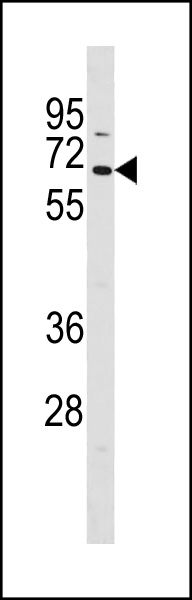
TCF7L1 Antibody (N-term)
Affinity Purified Rabbit Polyclonal Antibody (Pab)
- SPECIFICATION
- CITATIONS
- PROTOCOLS
- BACKGROUND

Application
| WB, E |
|---|---|
| Primary Accession | Q9HCS4 |
| Reactivity | Human |
| Host | Rabbit |
| Clonality | Polyclonal |
| Isotype | Rabbit IgG |
| Calculated MW | 62631 Da |
| Antigen Region | 70-98 aa |
| Gene ID | 83439 |
|---|---|
| Other Names | Transcription factor 7-like 1, HMG box transcription factor 3, TCF-3, TCF7L1, TCF3 |
| Target/Specificity | This TCF7L1 antibody is generated from rabbits immunized with a KLH conjugated synthetic peptide between 70-98 amino acids from the N-terminal region of human TCF7L1. |
| Dilution | WB~~1:1000 E~~Use at an assay dependent concentration. |
| Format | Purified polyclonal antibody supplied in PBS with 0.09% (W/V) sodium azide. This antibody is purified through a protein A column, followed by peptide affinity purification. |
| Storage | Maintain refrigerated at 2-8°C for up to 2 weeks. For long term storage store at -20°C in small aliquots to prevent freeze-thaw cycles. |
| Precautions | TCF7L1 Antibody (N-term) is for research use only and not for use in diagnostic or therapeutic procedures. |
| Name | TCF7L1 |
|---|---|
| Synonyms | TCF3 |
| Function | Participates in the Wnt signaling pathway. Binds to DNA and acts as a repressor in the absence of CTNNB1, and as an activator in its presence. Necessary for the terminal differentiation of epidermal cells, the formation of keratohyalin granules and the development of the barrier function of the epidermis (By similarity). Down-regulates NQO1, leading to increased mitomycin c resistance. |
| Cellular Location | Nucleus. |
| Tissue Location | Detected in hair follicles and skin keratinocytes, and at lower levels in stomach epithelium |

Thousands of laboratories across the world have published research that depended on the performance of antibodies from Abcepta to advance their research. Check out links to articles that cite our products in major peer-reviewed journals, organized by research category.
info@abcepta.com, and receive a free "I Love Antibodies" mug.

Provided below are standard protocols that you may find useful for product applications.
Background
Participates in the Wnt signaling pathway. Binds to DNA and acts as a repressor in the absence of CTNNB1, and as an activator in its presence. Necessary for the terminal differentiation of epidermal cells, the formation of keratohyalin granules and the development of the barrier function of the epidermis (By similarity). Down-regulates NQO1, leading to increased mitomycin c resistance.
If you have used an Abcepta product and would like to share how it has performed, please click on the "Submit Review" button and provide the requested information. Our staff will examine and post your review and contact you if needed.
If you have any additional inquiries please email technical services at tech@abcepta.com.
Ordering Information
Other Products
Shipping Information
