


 Foundational characteristics of cancer include proliferation, angiogenesis, migration, evasion of apoptosis, and cellular immortality. Find key markers for these cellular processes and antibodies to detect them.
 The SUMOplot™ Analysis Program predicts and scores sumoylation sites in your protein. SUMOylation is a post-translational modification involved in various cellular processes, such as nuclear-cytosolic transport, transcriptional regulation, apoptosis, protein stability, response to stress, and progression through the cell cycle.
 The Autophagy Receptor Motif Plotter predicts and scores autophagy receptor binding sites in your protein. Identifying proteins connected to this pathway is critical to understanding the role of autophagy in physiological as well as pathological processes such as development, differentiation, neurodegenerative diseases, stress, infection, and cancer.
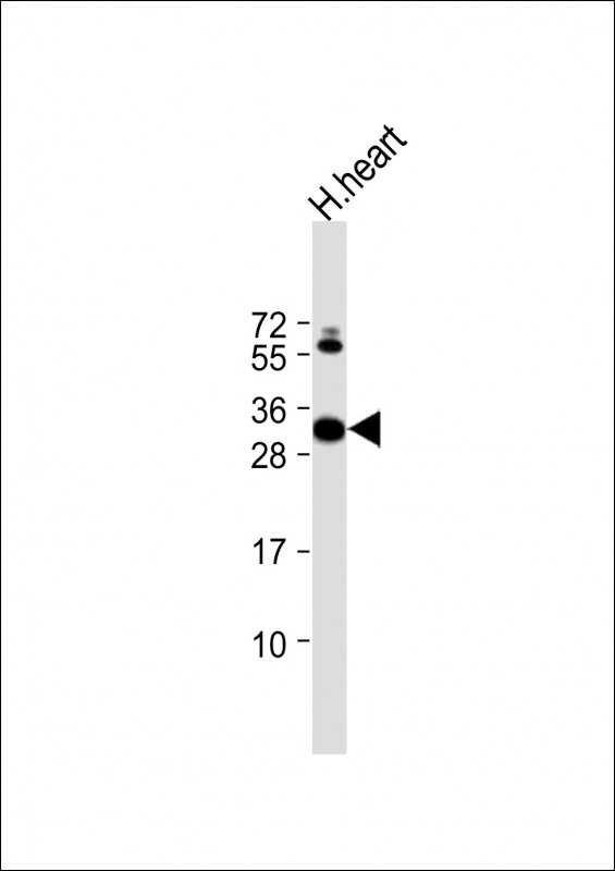
CLIC2 Antibody (Center)
Purified Rabbit Polyclonal Antibody (Pab)
- SPECIFICATION
- CITATIONS
- PROTOCOLS
- BACKGROUND

Application
| WB, E |
|---|---|
| Primary Accession | O15247 |
| Other Accession | Q5M883 |
| Reactivity | Human |
| Predicted | Rat |
| Host | Rabbit |
| Clonality | polyclonal |
| Isotype | Rabbit IgG |
| Calculated MW | 28356 Da |
| Gene ID | 1193 |
|---|---|
| Other Names | Chloride intracellular channel protein 2, XAP121, CLIC2 |
| Target/Specificity | This CLIC2 antibody is generated from a rabbit immunized with a KLH conjugated synthetic peptide between 105-137 amino acids from the Central region of human CLIC2. |
| Dilution | WB~~1:2000 E~~Use at an assay dependent concentration. |
| Format | Purified polyclonal antibody supplied in PBS with 0.09% (W/V) sodium azide. This antibody is purified through a protein A column, followed by peptide affinity purification. |
| Storage | Maintain refrigerated at 2-8°C for up to 2 weeks. For long term storage store at -20°C in small aliquots to prevent freeze-thaw cycles. |
| Precautions | CLIC2 Antibody (Center) is for research use only and not for use in diagnostic or therapeutic procedures. |
| Name | CLIC2 {ECO:0000303|PubMed:9339381, ECO:0000312|HGNC:HGNC:2063} |
|---|---|
| Function | In the soluble state, catalyzes glutaredoxin-like thiol disulfide exchange reactions with reduced glutathione as electron donor. Displays weak glutathione peroxidase activity (Probable) (PubMed:25581026). Can insert into membranes and form chloride ion channels. Membrane insertion seems to be redox-regulated and may occur only under oxidizing conditions. Modulates the activity of RYR2 and inhibits calcium influx. |
| Cellular Location | Cytoplasm. Membrane; Single-pass membrane protein. Note=Exists both as soluble cytoplasmic protein and as membrane protein with probably a single transmembrane domain |
| Tissue Location | Expressed in adult and fetal brain, heart, skeletal muscle, liver, lung, and spleen. Detected in adult stomach and testis Expressed in fetal thymus and kidney. |

Thousands of laboratories across the world have published research that depended on the performance of antibodies from Abcepta to advance their research. Check out links to articles that cite our products in major peer-reviewed journals, organized by research category.
info@abcepta.com, and receive a free "I Love Antibodies" mug.

Provided below are standard protocols that you may find useful for product applications.
Background
Can insert into membranes and form chloride ion channels. Channel activity depends on the pH. Membrane insertion seems to be redox-regulated and may occur only under oxydizing conditions. Modulates the activity of RYR2 and inhibits calcium influx.
References
Heiss N.S.,et al.Genomics 45:224-228(1997).
Ota T.,et al.Nat. Genet. 36:40-45(2004).
Ross M.T.,et al.Nature 434:325-337(2005).
Mural R.J.,et al.Submitted (SEP-2005) to the EMBL/GenBank/DDBJ databases.
Fan L.,et al.FEBS Lett. 540:77-80(2003).
If you have used an Abcepta product and would like to share how it has performed, please click on the "Submit Review" button and provide the requested information. Our staff will examine and post your review and contact you if needed.
If you have any additional inquiries please email technical services at tech@abcepta.com.
Ordering Information
Other Products
Shipping Information
