


 Foundational characteristics of cancer include proliferation, angiogenesis, migration, evasion of apoptosis, and cellular immortality. Find key markers for these cellular processes and antibodies to detect them.
 The SUMOplot™ Analysis Program predicts and scores sumoylation sites in your protein. SUMOylation is a post-translational modification involved in various cellular processes, such as nuclear-cytosolic transport, transcriptional regulation, apoptosis, protein stability, response to stress, and progression through the cell cycle.
 The Autophagy Receptor Motif Plotter predicts and scores autophagy receptor binding sites in your protein. Identifying proteins connected to this pathway is critical to understanding the role of autophagy in physiological as well as pathological processes such as development, differentiation, neurodegenerative diseases, stress, infection, and cancer.
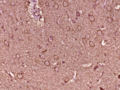
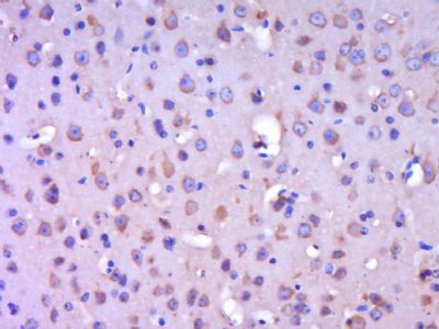
SPEF1 Polyclonal Antibody
Purified Rabbit Polyclonal Antibody (Pab)
- SPECIFICATION
- CITATIONS
- PROTOCOLS
- BACKGROUND

Application
| IHC-P, IHC-F, IF, ICC, E |
|---|---|
| Primary Accession | Q9Y4P9 |
| Reactivity | Rat, Pig, Bovine |
| Host | Rabbit |
| Clonality | Polyclonal |
| Calculated MW | 26987 Da |
| Gene ID | 25876 |
|---|---|
| Other Names | Sperm flagellar protein 1, SPEF1, C20orf28 |
| Dilution | IHC-P=1:100-500,IHC-F=1:100-500,ICC=1:100-500,IF=1:100-500,ELISA=1:5000-10000 |
| Format | 0.01M TBS(pH7.4), 0.09% (W/V) sodium azide and 50% Glyce |
| Storage | Store at -20 ℃ for one year. Avoid repeated freeze/thaw cycles. When reconstituted in sterile pH 7.4 0.01M PBS or diluent of antibody the antibody is stable for at least two weeks at 2-4 ℃. |
| Name | SPEF1 |
|---|---|
| Synonyms | C20orf28 |
| Function | Microtubule-associated protein involved in the stabilization of microtubules along the axis of migration during radial intercalation. Promotes the establishment and stabilization of an axis of microtubules required for the active migration of cells into the outer epithelium (By similarity). Microtubule-associated protein that promotes microtubule bundling and stabilizes microtubules against depolymerization in response to cold shock (By similarity). Essential for ciliary central apparatus formation which requires both its microtubule-binding and bundling activities and for ciliary localization of HYDIN and SPAG6 in ependymal cilia (By similarity). Binds actin in intestinal epithelial cells (IECs), essential for IECs survival and contributes to formation of filopodia and lamellipodia in migrating IECs (PubMed:31473225). Regulates planar cell polarity signaling pathway and asymmetric microtubule accumulation in ciliated epithelia (By similarity). |
| Cellular Location | Cytoplasm. Cell projection, cilium, flagellum {ECO:0000250|UniProtKB:Q99JL1} Cytoplasm, cytoskeleton, cilium axoneme {ECO:0000250|UniProtKB:Q0IH24} Apical cell membrane. Basolateral cell membrane. Cytoplasm, cytoskeleton, stress fiber. Cell projection, microvillus. Cell projection, lamellipodium. Cell projection, filopodium. Note=Present in the tails of developing and epididymal sperm, internal to the fibrous sheath and around the outer dense fibers of the sperm flagellum. Also found at the apical tip of cilia (By similarity). Colocalizes with TJP1 and CGN at sites of cell-cell contact in intestinal epithelial cells (PubMed:31473225) {ECO:0000250|UniProtKB:Q0IH24, ECO:0000269|PubMed:31473225} |
| Tissue Location | Expressed in the intestinal epithelial cells (at protein level). |
Research Areas
Citations (0)

Thousands of laboratories across the world have published research that depended on the performance of antibodies from Abcepta to advance their research. Check out links to articles that cite our products in major peer-reviewed journals, organized by research category.
Submit your citation using an Abcepta antibody to
info@abcepta.com, and receive a free "I Love Antibodies" mug.
info@abcepta.com, and receive a free "I Love Antibodies" mug.

Application Protocols
Provided below are standard protocols that you may find useful for product applications.
Abcepta welcomes feedback from its customers.
If you have used an Abcepta product and would like to share how it has performed, please click on the "Submit Review" button and provide the requested information. Our staff will examine and post your review and contact you if needed.
If you have any additional inquiries please email technical services at tech@abcepta.com.
$ 385.00
Cat# AP56775
Ordering Information
United States
AlbaniaAustraliaAustriaBelgiumBosnia & HerzegovinaBrazilBulgariaCanadaCentral AmericaChinaCroatiaCyprusCzech RepublicDenmarkEstoniaFinlandFranceGermanyGreeceHong KongHungaryIcelandIndiaIndonesiaIrelandIsraelItalyJapanLatviaLithuaniaLuxembourgMacedoniaMalaysiaMaltaMexicoNetherlandsNew ZealandNorwayPakistanPolandPortugalRomaniaSerbiaSingaporeSlovakiaSloveniaSouth AfricaSouth KoreaSpainSwedenSwitzerlandTaiwanTurkeyUnited KingdomUnited StatesVietnamWorldwideOthers
USA Headquarters
(888) 735-7227 / (858) 622-0099 or (858) 875-1900
Shipping Information
Domestic orders (in stock items)
Shipped out the same day. Orders placed after 1 PM (PST) will ship out the next business day.
International orders
Contact your local distributors
