


 Foundational characteristics of cancer include proliferation, angiogenesis, migration, evasion of apoptosis, and cellular immortality. Find key markers for these cellular processes and antibodies to detect them.
 The SUMOplot™ Analysis Program predicts and scores sumoylation sites in your protein. SUMOylation is a post-translational modification involved in various cellular processes, such as nuclear-cytosolic transport, transcriptional regulation, apoptosis, protein stability, response to stress, and progression through the cell cycle.
 The Autophagy Receptor Motif Plotter predicts and scores autophagy receptor binding sites in your protein. Identifying proteins connected to this pathway is critical to understanding the role of autophagy in physiological as well as pathological processes such as development, differentiation, neurodegenerative diseases, stress, infection, and cancer.
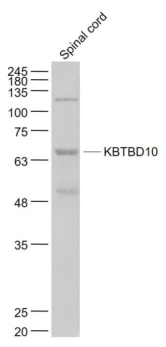
KBTBD10 Polyclonal Antibody
Purified Rabbit Polyclonal Antibody (Pab)
- SPECIFICATION
- CITATIONS
- PROTOCOLS
- BACKGROUND

Application
| WB, IHC-P, IHC-F, IF, E |
|---|---|
| Primary Accession | O60662 |
| Reactivity | Rat, Pig, Dog, Bovine |
| Host | Rabbit |
| Clonality | Polyclonal |
| Calculated MW | 68 KDa |
| Physical State | Liquid |
| Immunogen | KLH conjugated synthetic peptide derived from human KBTBD10 |
| Epitope Specificity | 542-606/606 |
| Purity | affinity purified by Protein A |
| Buffer | 0.01M TBS (pH7.4) with 1% BSA, 0.02% Proclin300 and 50% Glycerol. |
| SUBCELLULAR LOCATION | Cytoplasm. Cytoplasm, cytoskeleton. Cell projection, pseudopodium. Cell projection, ruffle. Note=Predominantly cytoplasmic but can co-localize with F-actin at the membrane ruffle-like structures at the tips of transformation-specific pseudopodia. |
| SIMILARITY | Contains 1 BTB (POZ) domain. Contains 5 Kelch repeats. |
| SUBUNIT | Interacts with NRAP (By similarity). Part of a complex that contains CUL3, RBX1 and KBTBD10. |
| Post-translational modifications | Ubiquitinated and probably targeted for proteasome-independent degradation. |
| Important Note | This product as supplied is intended for research use only, not for use in human, therapeutic or diagnostic applications. |
| Background Descriptions | Sarcosin contains 1 BTB (POZ) domain and is required for pseudopod elongation in transformed cells. Sarcosin mRNA is up-regulated by less than two folds in the heart in human patients with HCM. |
| Gene ID | 10324 |
|---|---|
| Other Names | Kelch-like protein 41, Kel-like protein 23, Kelch repeat and BTB domain-containing protein 10, Kelch-related protein 1, Sarcosin, KLHL41, KBTBD10, KRP1 |
| Dilution | WB=1:500-2000,IHC-P=1:100-500,IHC-F=1:100-500,IF=1:100-500,ELISA=1:5000-10000 |
| Storage | Store at -20 ℃ for one year. Avoid repeated freeze/thaw cycles. When reconstituted in sterile pH 7.4 0.01M PBS or diluent of antibody the antibody is stable for at least two weeks at 2-4 ℃. |
| Name | KLHL41 |
|---|---|
| Synonyms | KBTBD10, KRP1 |
| Function | Involved in skeletal muscle development and differentiation. Regulates proliferation and differentiation of myoblasts and plays a role in myofibril assembly by promoting lateral fusion of adjacent thin fibrils into mature, wide myofibrils. Required for pseudopod elongation in transformed cells. |
| Cellular Location | Cytoplasm. Cytoplasm, cytoskeleton {ECO:0000250|UniProtKB:A2AUC9}. Cell projection, pseudopodium {ECO:0000250|UniProtKB:Q9ER30}. Cell projection, ruffle {ECO:0000250|UniProtKB:Q9ER30}. Cytoplasm, myofibril, sarcomere, M line {ECO:0000250|UniProtKB:A2AUC9} Sarcoplasmic reticulum membrane Endoplasmic reticulum membrane Note=Predominantly cytoplasmic but can colocalize with F-actin at the membrane ruffle-like structures at the tips of transformation-specific pseudopodia. |
| Tissue Location | Sarcomeric muscle. |
Research Areas
Citations (0)

Thousands of laboratories across the world have published research that depended on the performance of antibodies from Abcepta to advance their research. Check out links to articles that cite our products in major peer-reviewed journals, organized by research category.
Submit your citation using an Abcepta antibody to
info@abcepta.com, and receive a free "I Love Antibodies" mug.
info@abcepta.com, and receive a free "I Love Antibodies" mug.

Application Protocols
Provided below are standard protocols that you may find useful for product applications.
Abcepta welcomes feedback from its customers.
If you have used an Abcepta product and would like to share how it has performed, please click on the "Submit Review" button and provide the requested information. Our staff will examine and post your review and contact you if needed.
If you have any additional inquiries please email technical services at tech@abcepta.com.
$ 385.00
Cat# AP58845
Ordering Information
United States
AlbaniaAustraliaAustriaBelgiumBosnia & HerzegovinaBrazilBulgariaCanadaCentral AmericaChinaCroatiaCyprusCzech RepublicDenmarkEstoniaFinlandFranceGermanyGreeceHong KongHungaryIcelandIndiaIndonesiaIrelandIsraelItalyJapanLatviaLithuaniaLuxembourgMacedoniaMalaysiaMaltaMexicoNetherlandsNew ZealandNorwayPakistanPolandPortugalRomaniaSerbiaSingaporeSlovakiaSloveniaSouth AfricaSouth KoreaSpainSwedenSwitzerlandTaiwanTurkeyUnited KingdomUnited StatesVietnamWorldwideOthers
USA Headquarters
(888) 735-7227 / (858) 622-0099 or (858) 875-1900
Other Products
Shipping Information
Domestic orders (in stock items)
Shipped out the same day. Orders placed after 1 PM (PST) will ship out the next business day.
International orders
Contact your local distributors
